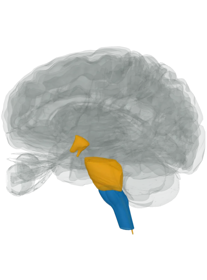
Made in: lit in blue
- Adrenergic cell groups C1, C2 and C3, in the medulla oblongata: C1 lies in the rostral and intermediate ventrolateral medulla, C2 in the rostral nucleus of the solitary tract, C3 in the rostral dorsomedial medulla. They are named for PNMT, the enzyme that turns noradrenaline into adrenaline; in rats C1 holds about 72% of these cells, C2 13% and C3 15%. [2][4]
Sent to: lit in orange
- Spinal cord (c1 → sympathetic preganglionic neurons): About a third of the C1 cells reach the preganglionic sympathetic neurons of the intermediolateral cell column. The atlas draws the cord only as its central canal, cropped just below the medulla, so that thin line is what is lit. [4]
- Paraventricular nucleus, lit as the hypothalamus (c1 → paraventricular nucleus): The C1 cells densely supply its parvocellular part. It is not a separate shape here, so the hypothalamus is lit. [4]
- Locus coeruleus, lit as the pons (c1 → locus coeruleus): Their input to the noradrenaline neurons of the locus coeruleus is dense and excitatory. It is not a separate shape here, so the pons is lit. [4]
- Also reached but inside parts already lit: the dorsal vagal complex and the raphe pallidus in the medulla and the parabrachial nuclei in the pons, which the C1 review lists among their other targets. [4]
- Not lit: the adrenal glands, outside the brain, which make most of the adrenaline that circulates in the body. Their medulla’s chromaffin cells release a mix of about 85% adrenaline and 15% noradrenaline. [3][6]
The pathways, as the sources name them
- C1 → sympathetic preganglionic neurons, from the presympathetic C1 cells down to the intermediolateral cell column of the spinal cord. These spinal neurons drive the sympathetic nervous system, including the adrenal medulla; the C1 cells help regulate it, particularly the heart and blood vessels. [4][7][3] See What part of the brain controls heart rate and blood pressure?
- C1 → paraventricular nucleus, from the C1 cells up to the paraventricular nucleus of the hypothalamus. Through these direct projections the C1 cells help regulate the hypothalamic–pituitary–adrenal axis. [4][7] See What part of the brain controls the stress response?
- C1 → locus coeruleus, from the C1 cells to the locus coeruleus in the upper pons. The brain’s main noradrenaline source is among the targets the C1 review lists, with dense, excitatory input. [4]
Good to know
- An honest limit: these cells are named adrenergic because they carry PNMT, so they are thought to release adrenaline, but the C1 review finds no direct evidence that they do; their fast messenger is glutamate. [2][4]
- One step from noradrenaline: the last step in making adrenaline is PNMT adding a methyl group to noradrenaline’s amine. [1]
- Adrenaline and epinephrine are the same molecule, and “adrenalin” is another spelling; it plays an essential role in the fight-or-flight response. [1]
- The C1 cells respond to stressors such as low blood sugar, low oxygen, pain and low blood pressure, and help set off the body’s response to them. [7] See What part of the brain controls the stress response?
- First from the adrenal gland: Napoleon Cybulski obtained adrenal extracts in 1895, and in 1901 Jokichi Takamine isolated and purified the hormone from the adrenal glands of sheep and oxen. [1]
What it takes part in
Each lit with every part that takes part: What part of the brain controls the stress response? · What part of the brain controls heart rate and blood pressure? · What part of the brain controls hormones?
Where it is made, each on its own page: Adrenergic cell groups C1, C2 and C3, in the medulla oblongata.