Where is it in the brain?
This page was written with AI.
Suggestions appear below as you type, grouped into parts and what they do; press the down arrow to reach them.
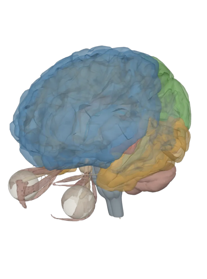
Loading the 3D brain…
Where is it? One page per structure
Together they make a brain map you can search: each page lights the structure up and slices through it, then says where it is, what it does and what it connects to, with every fact cited.
- Where is the hippocampus? Deep in the inner (medial) part of each temporal lobe, curled along the floor of the lateral ventricle’s lower (temporal) horn.
- Where is the amygdala? An almond-shaped cluster of nuclei deep in the medial temporal lobe of each hemisphere, just in front of the hippocampus.
- Where is the cerebellum? At the back of the head, under the occipital lobes and behind the pons and medulla, separated from the cerebrum by a sheet of dura (the tentorium cerebelli).
- Where is the thalamus? Two egg-shaped masses of grey matter in the centre of the brain, one each side of the third ventricle, sitting on top of the brainstem.
- Where is the hypothalamus? At the base of the brain just below the thalamus, forming the floor and lower walls of the third ventricle; the pituitary gland hangs from it on a stalk.
- Where is the brainstem? The stalk joining the cerebrum to the spinal cord, in front of the cerebellum: midbrain at the top, pons in the middle, medulla oblongata at the bottom.
- Where is the frontal lobe? The front of each cerebral hemisphere, ahead of the central sulcus and above the lateral (Sylvian) fissure — the largest of the four lobes.
- Where is the temporal lobe? The lower side of each hemisphere, below the lateral (Sylvian) fissure, behind the temple and above the ear.
- Where is the occipital lobe? The back of each cerebral hemisphere, behind the parietal and temporal lobes and above the cerebellum.
- Where is the prefrontal cortex? The front part of the frontal lobe, behind the forehead and ahead of the motor and premotor areas.
- Where is the parietal lobe? The upper middle of each hemisphere, behind the central sulcus, above the temporal lobe and in front of the occipital lobe.
- Where is the insula? Cortex folded deep inside the lateral (Sylvian) fissure of each hemisphere, hidden under the frontal, parietal and temporal lobes.
- Where is the cingulate gyrus? On the inner (medial) face of each hemisphere, arching over the top of the corpus callosum from front to back.
- Where is the corpus callosum? A broad arched band of white matter at the bottom of the deep groove between the two hemispheres, roofing the lateral ventricles.
- Where is the pituitary gland? A pea-sized gland under the base of the brain, sitting in a saddle-shaped pocket of the sphenoid bone (the sella turcica), attached to the hypothalamus by a stalk.
- Where is the pineal gland? A small pine-cone-shaped gland on the midline at the back of the third ventricle, just above the superior colliculi of the midbrain.
- Where is the midbrain? The top section of the brainstem, just below the thalamus and above the pons; its back surface carries four small bumps, the colliculi.
- Where is the pons? The bulging middle section of the brainstem, between the midbrain above and the medulla below, directly in front of the cerebellum.
- Where is the medulla oblongata? The lowest section of the brainstem, below the pons, continuing into the spinal cord at the large opening in the base of the skull (foramen magnum).
- Where is the caudate nucleus? A C-shaped nucleus deep in each hemisphere, curving along the outer wall of the lateral ventricle; part of the basal ganglia.
- Where is the putamen? A rounded nucleus deep in each hemisphere, beneath the insula and outside the globus pallidus; part of the basal ganglia.
- Where is the globus pallidus? Deep in each hemisphere, just inside the putamen and outside the internal capsule; part of the basal ganglia.
- Where is the motor cortex (precentral gyrus)? A strip of cortex over the top and side of each hemisphere, at the back edge of the frontal lobe, just in front of the central sulcus.
- Where is the somatosensory cortex (postcentral gyrus)? A strip of cortex just behind the central sulcus, the front edge of the parietal lobe, running from the top of the head down the side.
- Where is the superior frontal gyrus? The top horizontal strip of each frontal lobe, running front to back along the upper edge beside the deep groove between the hemispheres.
- Where is the middle frontal gyrus? The middle horizontal strip on the outer side of each frontal lobe, between the superior and inferior frontal gyri.
- Where is the inferior frontal gyrus? The lowest horizontal strip on the outer side of each frontal lobe, just above the lateral (Sylvian) fissure.
- Where is the orbitofrontal cortex? The underside of each frontal lobe, resting on the bony roofs of the eye sockets (orbits); on this model its orbital gyri.
- Where is Broca’s area? The back part of the inferior frontal gyrus (pars opercularis and pars triangularis), in the frontal lobe just above the lateral fissure, usually on the left.
- Where is Wernicke’s area? The posterior (back) part of the superior temporal gyrus, Brodmann area 22, in the dominant hemisphere, typically the left.
- Where is the auditory cortex? The upper side of each temporal lobe: the superior temporal gyrus where it runs into the lateral sulcus as the transverse temporal (Heschl’s) gyri. On both sides.
- Where is the visual cortex? The back of each occipital lobe: the primary visual cortex lies in and around the calcarine sulcus on the lobe’s inner face and at its back tip, the occipital pole. On both sides.
- Where is the premotor cortex? The back of each frontal lobe’s outer side, just in front of the primary motor cortex: the lateral part of Brodmann area 6, on the rear parts of the superior and middle frontal gyri. On both sides.
- Where is the supplementary motor area? On the inner, midline face at the top of each frontal lobe, just in front of the leg part of the primary motor cortex: the medial part of Brodmann area 6, on the superior frontal gyrus. On both sides.
- Where is the entorhinal cortex? On the inner underside of each temporal lobe: the front (rostral) part of the parahippocampal gyrus, beside the hippocampus. On both sides.
- Where are the frontal eye fields? In the frontal lobe, one on each side: around where the back of the middle frontal gyrus meets the precentral gyrus, inside Brodmann area 8, just in front of the premotor cortex.
- Where is the temporal pole? The front tip of each temporal lobe: Brodmann area 38, mostly on the most forward parts of the superior and middle temporal gyri. On both sides.
- Where is the gustatory cortex? Deep in the side of each hemisphere: the front of the insula plus the frontal operculum of the inferior frontal gyrus, the lid of cortex that folds over it. On both sides.
- Where is the piriform cortex? On the inner underside of each temporal lobe, at its front: the cortical amygdala, the uncus and the front of the parahippocampal gyrus, between the insula and the temporal lobe. On both sides.
- Where is the precuneus? On the inner (medial) face of each parietal lobe, toward the back: in front of the cuneus, behind the paracentral lobule and above the posterior cingulate. It is the inner part of the superior parietal lobule.
- Where is the cuneus? On the inner (medial) face of each occipital lobe: the wedge between the parieto-occipital sulcus in front and the calcarine sulcus below.
- Where is the lingual gyrus? On the inner underside of each occipital lobe, below the calcarine sulcus; it runs back to the occipital pole and forward to join the parahippocampal gyrus.
- Where is the uncus? The hooked front end of the parahippocampal gyrus, on the inner underside of each temporal lobe, just behind its tip.
- Where are the opercula? Around the lateral sulcus on the side of each hemisphere: the edges of the frontal, parietal and temporal lobes that fold over the insula and hide it.
- Where is the anterior cingulate cortex? On the inner (medial) face of each hemisphere: the front part of the cingulate gyrus, curving like a collar round the front of the corpus callosum. Brodmann areas 24, 32 and 33.
- Where is the posterior cingulate cortex? On the inner (medial) face of each hemisphere: the back part of the cingulate gyrus, behind the anterior cingulate cortex and above the corpus callosum. Brodmann areas 23 and 31.
- Where is the dorsolateral prefrontal cortex? On the upper outer side of each frontal lobe, toward the front: in the middle frontal gyrus, the lateral parts of Brodmann areas 9 and 46.
- Where is the ventromedial prefrontal cortex? The lower (ventral), middle (medial) part of the prefrontal cortex, at the bottom of the frontal lobes where the two halves face each other; the medial orbitofrontal cortex is its lowest part.
- Where is the paracentral lobule? At the top of the inner (medial) face of each hemisphere, where the precentral and postcentral gyri continue over the upper edge: between the paracentral sulcus in front and the marginal sulcus behind, above the cingulate sulcus.
- Where is the gyrus rectus? On the underside of each frontal lobe, the strip right beside the midline (the longitudinal fissure), on the inner side of the olfactory sulcus where the olfactory bulb and tract lie.
- Where are the pars opercularis, pars triangularis and pars orbitalis? Along the inferior frontal gyrus, low on the side of the frontal lobe, from back to front: the pars opercularis, the pars triangularis and the pars orbitalis, divided by two small branches of the lateral sulcus.
- Where are the rostrum, genu, body and splenium of the corpus callosum? Along the corpus callosum on the midline, front to back: the rostrum underneath at the front, the genu where it bends at the front, the body (trunk) along the top, and the splenium, the thick back end.
- Where is the fusiform gyrus? On the underside of each temporal and occipital lobe, between the parahippocampal gyrus on the inside and the inferior temporal gyrus on the outside.
- Where is the parahippocampal gyrus? On the inner underside of each temporal lobe, running alongside and below the hippocampus.
- Where is the internal capsule? A band of white matter deep in each hemisphere, between the caudate nucleus and thalamus on the inside and the putamen and globus pallidus on the outside.
- Where are the ventricles of the brain? Four connected fluid-filled spaces deep inside the brain: a lateral ventricle in each hemisphere, the third ventricle on the midline between the two thalami, and the fourth ventricle between the brainstem and the cerebellum.
- Where are the lateral ventricles? Two C-shaped fluid-filled cavities, one inside each cerebral hemisphere, curving from the frontal lobe back and down into the temporal lobe.
- Where is the third ventricle? A narrow fluid-filled slit on the midline between the left and right thalamus, with the hypothalamus forming its floor.
- Where is the fourth ventricle? A tent-shaped fluid-filled space between the pons and upper medulla in front and the cerebellum behind.
- Where are the basal ganglia? Deep inside each cerebral hemisphere, at the base of the forebrain: the caudate nucleus curving beside the lateral ventricle, and the putamen and globus pallidus further out, with the internal capsule running between them.
- Where is the limbic system? On the inner edge of each hemisphere, curving around the thalamus and the top of the brainstem: the cingulate gyrus above, the hippocampus, parahippocampal gyrus and amygdala in the inner temporal lobe below, the fornix arching between them, and the hypothalamus with its mammillary bodies at the base.
- Where is the diencephalon? In the middle of the brain, between the cerebral hemispheres and the midbrain, wrapped around the third ventricle: the thalamus above, the hypothalamus below, the pineal gland at the back.
- Where is the epithalamus? The back (dorsal) part of the diencephalon, at the back of the roof of the third ventricle, above the midbrain: the habenula and stria medullaris on the top inner edge of each thalamus, the pineal gland and posterior commissure on the midline.
- Where is the inferior parietal lobule? The lower half of each parietal lobe, below the intraparietal sulcus and behind the postcentral sulcus: the supramarginal gyrus in front, curving over the end of the lateral fissure, and the angular gyrus behind it.
- Where is the forebrain? The front and top of the brain, its largest division: both cerebral hemispheres (the cerebrum) and, at their core, the diencephalon above the midbrain.
- Where is the hindbrain? The back and bottom of the brain, below the midbrain: the pons and medulla oblongata (the lower brainstem) and the cerebellum behind them.
- Where is the cerebrum? The top and front of the brain, above the brainstem and in front of the cerebellum: the two cerebral hemispheres, split by the longitudinal fissure. Lit here: their cortex, as the five lobes of each side.
- Where are the cerebral hemispheres? The left and right halves of the cerebrum, side by side on top of the brainstem, split by the longitudinal fissure and joined across it by the corpus callosum.
- Where is the central sulcus? A deep groove across the side of each hemisphere, between the precentral gyrus in front and the postcentral gyrus behind; it separates the frontal lobe from the parietal lobe.
- Where is the lateral sulcus (Sylvian fissure)? A deep groove on the side of each hemisphere, with the frontal and parietal lobes above it and the temporal lobe below; the insula lies folded in its depth.
- Where is the longitudinal fissure? The deep groove down the middle of the brain, from front to back, between the left and right cerebral hemispheres; the corpus callosum lies at its floor.
- Where is the parieto-occipital sulcus? Mostly on the inner (medial) face of each hemisphere, running down and forward from the top edge to meet the calcarine sulcus; only a short notch shows on the outer surface.
- Where is the calcarine sulcus? On the inner (medial) face of the occipital lobe at the back of each hemisphere, running forward from near the occipital pole to below the back end of the corpus callosum.
- Where is the cingulate sulcus? On the inner (medial) face of each hemisphere, arching over the corpus callosum between the cingulate gyrus below and the frontal and parietal lobes above.
- Where is the superior temporal sulcus? A long groove running front to back along the outer side of each temporal lobe, below the lateral sulcus, between the superior temporal gyrus above and the middle temporal gyrus below.
- Where is the intraparietal sulcus? A groove on the outer side of each parietal lobe, between the superior parietal lobule above and the inferior parietal lobule (the supramarginal and angular gyri) below.
- Where is the collateral sulcus? A long groove on the underside of each hemisphere, from near the back tip almost to the front tip of the temporal lobe; in front it runs between the parahippocampal gyrus on the inner side and the fusiform gyrus on the outer side.
- Where is the precentral sulcus? A groove on the outer side of each frontal lobe, parallel to and in front of the central sulcus, between the precentral gyrus behind and the superior, middle and inferior frontal gyri in front.
- Where is the postcentral sulcus? A groove on the outer side of each parietal lobe, parallel to and behind the central sulcus, between the postcentral gyrus in front and the superior parietal lobule and supramarginal gyrus behind.
- Where is the superior frontal sulcus? A groove running front to back along the top of each frontal lobe, from near the precentral sulcus almost to the frontal pole, between the superior frontal gyrus above (towards the midline) and the middle frontal gyrus below.
- Where is the inferior frontal sulcus? A groove running front to back on the outer side of each frontal lobe, between the middle frontal gyrus above and the inferior frontal gyrus below; at the back it meets the inferior precentral sulcus.
- Where is the occipitotemporal sulcus? A long groove on the underside of each hemisphere, from near the back tip of the brain almost to the front tip of the temporal lobe, between the inferior temporal gyrus on the outer side and the fusiform gyrus on the inner side.
- Where is the angular gyrus? In the lower (inferior) parietal lobule of each hemisphere, behind the supramarginal gyrus, near where the parietal, temporal and occipital lobes meet.
- Where is the supramarginal gyrus? In the lower (inferior) parietal lobule of each hemisphere, arching over the upturned back end of the lateral (Sylvian) fissure.
- Where is the superior parietal lobule? The upper part of each parietal lobe, behind the postcentral gyrus and above the intraparietal sulcus.
- Where is the superior temporal gyrus? The top strip of each temporal lobe, running along the lower edge of the lateral (Sylvian) fissure above the ear.
- Where is the middle temporal gyrus? The middle horizontal strip on the outer side of each temporal lobe, between the superior and inferior temporal sulci.
- Where is the inferior temporal gyrus? The lowest strip on the outer side of each temporal lobe, curving round onto its underside above the cerebellum.
- Where is the fornix? A C-shaped bundle of white matter on each side that arches from the hippocampus in the temporal lobe up and forward under the corpus callosum, then curves down in front of the thalamus to the mammillary bodies.
- Where is the anterior commissure? A small bundle of white fibres crossing the midline in front of the columns of the fornix, at the front of the third ventricle.
- Where is the posterior commissure? A rounded band of white fibres crossing the midline on the back (dorsal) side of the front end of the cerebral aqueduct.
- Where is the septum (septum of telencephalon)? On the midline under the front of the corpus callosum, between the front horns of the two lateral ventricles and down to the fornix: the septal area. The atlas names this shape only "septum of telencephalon".
- Where are the cerebral peduncles? The two thick stalks on the front of the midbrain, one on each side, running down from the cerebrum into the pons.
- Where are the mammillary bodies? Two small round bumps on the underside of the brain, side by side at the back of the hypothalamus, just in front of the midbrain.
- Where is the habenula? A small paired nucleus at the top back of the thalamus on each side, beside the stalk of the pineal gland, part of the epithalamus.
- Where is the superior colliculus? The upper pair of the four small bumps (colliculi) on the back roof of the midbrain, just below the pineal gland and behind the cerebral aqueduct.
- Where is the inferior colliculus? The lower pair of the four small bumps on the back roof of the midbrain, below the superior colliculi and just above the top of the cerebellum.
- Where is the lateral geniculate body? A small knee-shaped bump on the underside of the back of each thalamus, where the optic tract ends.
- Where is the medial geniculate body? A small oval bump on the underside of the back of each thalamus, just inside the lateral geniculate body, linked to the inferior colliculus below.
- Where is the cerebral aqueduct? A narrow canal through the midbrain, about 15 mm long, running from the back of the third ventricle down to the fourth ventricle, with the colliculi behind it.
- Where is the optic chiasm? An X-shaped crossing on the underside of the brain, where the two optic nerves meet, just below the hypothalamus and in front of the stalk of the pituitary gland.
- Where is the optic nerve? Runs from the back of each eye, back through the eye socket and the optic canal of the skull, to the optic chiasm under the front of the brain.
- Where is the optic tract? A flat band on each side running back from the optic chiasm around the cerebral peduncle of the midbrain to the lateral geniculate body of the thalamus.
- Where is the oculomotor nerve? Leaves the front of the midbrain and runs forward into each eye socket; this atlas draws only its two end branches inside the eye socket (superior and inferior), so those are what is lit here.
- Where is the trochlear nerve? The cranial nerve with the fewest fibres and the only one to leave the back of the brainstem: from just below the inferior colliculi it winds forward round the midbrain to the eye socket.
- Where is the vagus nerve? Leaves the side of the medulla oblongata, behind the olive; exits the skull through the jugular foramen and runs down the neck into the chest and abdomen.
- Where is the trigeminal nerve? Leaves the side of the pons at mid-pons level; its three divisions exit the skull through the superior orbital fissure, the foramen rotundum and the foramen ovale.
- Where is the facial nerve? Leaves the brainstem at the border of the pons and the medulla, enters the temporal bone through the internal acoustic meatus and exits the skull through the stylomastoid foramen.
- Where is the vestibulocochlear nerve? Leaves the brainstem at the border of the pons and the medulla, in the cerebellopontine angle; runs into the internal acoustic meatus to the inner ear.
- Where is the abducens nerve? Leaves the brainstem at the border of the pons and the medulla, close to the midline; climbs the clivus through Dorello’s canal and the cavernous sinus and enters the eye socket through the superior orbital fissure.
- Where is the glossopharyngeal nerve? Leaves the side of the upper medulla, between the olive and the inferior cerebellar peduncle, just above the vagus; exits the skull through the jugular foramen.
- Where is the accessory nerve? Its spinal part rises from the upper spinal cord (from where the cord meets the medulla down to about C6), enters the skull through the foramen magnum and leaves through the jugular foramen; a small cranial part comes from the medulla.
- Where is the hypoglossal nerve? Leaves the front of the medulla as rootlets in the groove between the pyramid and the olive; exits the skull through the hypoglossal canal of the occipital bone.
- Where are the extraocular muscles? Six per eye inside the eye socket: four rectus muscles from a tendon ring at the back of the socket to the eyeball, and two oblique muscles.
- Where is the eyeball? In the front of each eye socket (orbit), below the front of the frontal lobe; the optic nerve leaves its back. Drawn here as its white outer coat, the sclera.
- Where is the spinal cord? The spinal cord continues the medulla oblongata down from the foramen magnum; this atlas draws it only as its central canal, the thin fluid channel down its middle, cropped about 6 mm below the medulla.
- Where is the substantia nigra? In the midbrain, just behind the cerebral peduncles: one band on each side of the midline.
- Where is the red nucleus? In the upper (rostral) midbrain, in the tegmentum next to the substantia nigra.
- Where is the nucleus accumbens? At the bottom of the striatum in the basal forebrain, under the internal capsule, where it merges with the caudate nucleus and putamen; one in each hemisphere.
- Where is the locus coeruleus? In the pons: at the back of its upper part, in the side of the floor of the fourth ventricle.
- Where is the suprachiasmatic nucleus? In the front of the hypothalamus, directly above the optic chiasm, one on each side of the third ventricle.
- Where is the ventral tegmental area? In the midbrain: a group of neurons close to the midline on its floor, between the two substantia nigra.
- Where is the periaqueductal grey? In the midbrain: the grey matter wrapped around the cerebral aqueduct, the canal that runs through it.
- Where are the raphe nuclei? Along the midline of the brainstem, as a seam of cell groups running through the midbrain, pons and medulla oblongata.
- Where is the olfactory bulb? On the underside of the frontal lobe, in the olfactory sulcus beside the midline, resting on the cribriform plate above the nose; one on each side.
- Where is the cerebellar vermis? The narrow strip along the midline of the cerebellum that joins its two hemispheres, at the back of the skull.
- Where are the cerebellar peduncles? Three paired fibre bundles joining the cerebellum to the back of the brainstem: the superior to the midbrain, the middle to the pons, the inferior to the medulla oblongata.
- Where are the cerebellar lobes? The cerebellum’s three lobes: the anterior lobe in front of the primary fissure, the much larger posterior lobe behind it and the small flocculonodular lobe, cut off by the posterolateral fissure.
- Where is the claustrum? Deep in each hemisphere, between the insula on the outside and the putamen on the inside, with the extreme and external capsules either side of it; one on each side.
- Where is the subthalamic nucleus? Just below the thalamus and above the substantia nigra at the top of the midbrain, on the inner side of the internal capsule; one on each side.
- Where is the inferior olive? Inside the medulla oblongata, under the olive: the oval bulge beside each pyramid on the medulla’s front surface; one on each side.
- Where is the superior olivary complex? Inside the lower pons, one on each side, beside the trapezoid body, the band where hearing fibres cross the midline.
- Where is the dentate nucleus? Deep in the white matter of each cerebellar hemisphere; the outermost and largest of the four pairs of deep cerebellar nuclei.
- Where are the deep cerebellar nuclei? Deep in the white matter at the core of the cerebellum, over the roof of the fourth ventricle; from the midline out: fastigial, interposed (globose + emboliform), dentate. One set on each side.
- Where is the dentate gyrus? Inside the hippocampus, deep in the temporal lobe: one of its subfields; one on each side. Only its tail, the band of Giacomini, shows on the surface.
- Where is the reticular formation? Through the core of the whole brainstem, from the lower end of the medulla oblongata up through the pons to the top of the midbrain.
- Where is the nucleus basalis? In the basal forebrain, under the globus pallidus and the anterior commissure and to the side of the front of the hypothalamus, in the substantia innominata; one on each side.
- Where is the paraventricular nucleus? In the hypothalamus, right beside the third ventricle, one on each side.
- Where is the supraoptic nucleus? At the base of the hypothalamus, next to the optic chiasm, one on each side.
- Where is the pulvinar? The back end of the thalamus, overhanging the superior colliculi of the midbrain; one on each side.
- Where is the ventral posterior nucleus? In the lower (ventral) tier of the outer part of the thalamus, towards its back; one on each side.
- Where is the anterior nucleus of the thalamus? The front end of the thalamus, its anterior group; one on each side.
- Where is the mediodorsal nucleus? The middle of the thalamus, near the midline, on the inner side of the internal medullary lamina; one on each side.
- Where is the reticular nucleus of the thalamus? A thin shell over the outer (lateral) side of each thalamus, between it and the internal capsule.
- Where are the ventral lateral and ventral anterior nuclei? The front part of the thalamus’s lower outer tier: the ventral anterior nucleus at the front, the ventral lateral nucleus behind it.
- Where are the intralaminar nuclei? Embedded in the Y-shaped internal medullary lamina, the white-matter sheet that splits each thalamus into its groups.
- Where is the arcuate nucleus? At the base of the hypothalamus, hugging the bottom of the third ventricle just above the median eminence, where the pituitary stalk leaves.
- Where is the lateral hypothalamus? The outer zone of each half of the hypothalamus, running its whole length front to back, away from the third ventricle.
- Where is the area postrema? A pair of small swellings on the back of the lower medulla oblongata, at the bottom tip of the fourth ventricle, just above the obex.
- Where is the solitary nucleus? A column of grey matter running lengthwise through the back and outer part of the medulla oblongata, around the solitary tract.
- Where is the dorsal vagal nucleus? In the medulla oblongata, just under the floor of the fourth ventricle, beside the hypoglossal nucleus.
- Where is the nucleus ambiguus? Deep in the outer part of the upper medulla oblongata, in the reticular formation, just behind the inferior olive.
- Where are the vestibular nuclei? Four nuclei on the floor of the fourth ventricle, spread across the pons and the medulla oblongata.
- Where are the cochlear nuclei? On the back and outer side of the brainstem, spanning the junction of the pons and the medulla oblongata.
- Where are the hippocampal fields (CA1–CA4 and the subiculum)? Inside the hippocampus, deep in each temporal lobe: CA3, CA2 and CA1 in a row along its curled band of cortex, CA4 tucked inside the dentate gyrus, then the subiculum.
- Where is the circle of Willis? A ring of linked arteries on the underside of the brain, in the hollow between the optic chiasm in front and the brainstem behind: the two internal carotids and the top of the basilar artery feed it.
- Where is the middle cerebral artery? The largest branch of the internal carotid: runs sideways under the brain, then up into the lateral (Sylvian) fissure over the insula, and fans out over the outer face of the hemisphere.
- Where is the anterior cerebral artery? Starts from the internal carotid under the front of the brain, runs forward above the optic nerve into the deep groove between the hemispheres, then arches back over the corpus callosum (as the pericallosal and callosomarginal arteries).
- Where is the posterior cerebral artery? The two end branches of the basilar artery, at the top of the brainstem: each curves back around the midbrain to the underside of the temporal lobe and the inner face of the occipital lobe.
- Where is the basilar artery? A single midline artery, about 3 cm long, lying in the groove on the front of the pons; formed where the two vertebral arteries join at the lower edge of the pons.
- Where is the internal carotid artery? Rises up each side of the neck, enters the skull through the carotid canal, runs through the cavernous sinus beside the pituitary and ends under the brain beside the optic chiasm; drawn here from the level of the foramen magnum up.
- Where is the vertebral artery? Climbs through the neck vertebrae, enters the skull through the foramen magnum and runs up the front of the medulla to join its partner at the lower edge of the pons; drawn here from the foramen magnum up (the part inside the skull).
- Where is the anterior communicating artery? A short cross-link, only a few millimetres long, between the left and right anterior cerebral arteries in front of the optic chiasm, at the front of the circle of Willis.
- Where is the posterior communicating artery? A short artery on each side of the circle of Willis, running back from the internal carotid to the posterior cerebral artery, beside the mammillary bodies and close to the oculomotor nerve.
- Where are the cerebellar arteries? Three arteries on each side, from top to bottom: the superior cerebellar from the top of the basilar, the anterior inferior cerebellar from its lower part, the posterior inferior cerebellar from the vertebral artery; each winds back around the brainstem to the cerebellum.
- Where is the choroid plexus? A strip of tissue inside each lateral ventricle, running through its body, atrium and lower (temporal) horn, but not its front or back horns.
- Where is the tentorium cerebelli? A fold of the dura mater stretched like a tent between the cerebellum below and the occipital lobes above; the atlas ships only its right half.
- Where is the stria terminalis? A thin fibre band on the floor of each lateral ventricle, on the line between the thalamus and the caudate nucleus, from the temporal horn round to the interventricular foramen.
- Where is the lamina terminalis? A thin sheet on the midline forming the front wall of the forebrain, from the interventricular foramen down to the base of the optic stalk, just in front of the tuber cinereum.
- Where are the interventricular foramina? Two short channels near the midline, one from each lateral ventricle down into the third ventricle, with the fornix in front and the thalamus behind.
- Where is the commissure of the fornix? A thin triangular sheet where the inner parts of the body of the fornix meet, under the back of the corpus callosum.
- Where is the stria medullaris? A horizontal ridge on the inner (medial) surface of each thalamus, along the roof of the third ventricle back to the habenula.
- Where is the white matter? The inner bulk of each cerebral hemisphere, under the grey cortex and around the deep grey nuclei and the ventricles; the corpus callosum joins the two halves.
- Where is the corona radiata? In the white matter of each hemisphere, above the basal ganglia: the internal capsule’s fibres fan out from it up towards the whole cortex; higher up it continues as the centrum semiovale.
- Where are the external and extreme capsules? Two thin sheets of white matter deep in each hemisphere, between the putamen and the insula: the external capsule next to the putamen, then the claustrum, then the extreme capsule next to the insula.
- Where is the basolateral amygdala? The deep nucleus group of each amygdala, in the inner temporal lobe: the lateral, basal and accessory basal nuclei, about half its volume.
- Where is the central amygdala? A small nucleus in the upper (dorsal) part of each amygdala, near where the stria terminalis leaves it; about 3% of the amygdala.
- Where is the bed nucleus of the stria terminalis? In the basal forebrain on each side, on top of the anterior commissure where it crosses, under the caudate nucleus, with the stria terminalis on its inner side.
- Where is the preoptic area? The front end of the hypothalamus, around the front of the third ventricle, between the anterior commissure above and the optic chiasm below.
- Where is the ventromedial hypothalamus? Low in the middle (tuberal) region of the hypothalamus, close to the midline, with the arcuate nucleus at its lower outer edge.
- Where is the dorsomedial hypothalamus? High in the middle (tuberal) region of the hypothalamus, near the midline, directly above the ventromedial nucleus.
What part of the brain does it? One page per function
Each page lights every part that takes part in one job at once, so you can turn the brain and see how they sit together, with each part’s role cited.
- What part of the brain controls balance? Cerebellum, Brainstem, Pons, Medulla oblongata, Midbrain, Thalamus, Insula, Occipital lobe
- What part of the brain controls memory? Hippocampus, Parahippocampal gyrus, Temporal lobe, Prefrontal cortex, Thalamus, Amygdala
- What part of the brain controls emotions? Amygdala, Hypothalamus, Cingulate gyrus, Insula, Prefrontal cortex
- What part of the brain controls movement? Precentral gyrus, Postcentral gyrus, Cerebellum, Caudate nucleus, Putamen, Globus pallidus, Internal capsule, Thalamus, Brainstem, Superior frontal gyrus, Middle frontal gyrus
- What part of the brain controls vision? Optic nerve, Optic chiasm, Optic tract, Lateral geniculate body, Occipital lobe, Temporal lobe, Parietal lobe, Fusiform gyrus, Superior colliculus
- What part of the brain controls sleep? Hypothalamus, Pineal gland, Pons, Thalamus, Brainstem
- What part of the brain controls consciousness? Midbrain, Pons, Medulla oblongata, Thalamus, Hypothalamus, Cingulate gyrus, Prefrontal cortex
- What part of the brain controls the bladder? Pons, Midbrain, Spinal cord (central canal), Insula, Cingulate gyrus, Prefrontal cortex, Hypothalamus
- What part of the brain controls breathing? Medulla oblongata, Pons, Brainstem, Precentral gyrus
- What part of the brain controls hunger? Hypothalamus, Medulla oblongata, Brainstem, Insula, Prefrontal cortex
- What part of the brain feels touch, temperature and pain? Postcentral gyrus, Thalamus, Parietal lobe, Insula, Cingulate gyrus, Brainstem
- What part of the brain controls speech and understands language? Inferior frontal gyrus, Superior temporal gyrus, Supramarginal gyrus, Angular gyrus, Precentral gyrus, Cerebellum, Thalamus
- What part of the brain controls hearing? Superior temporal gyrus, Medial geniculate body, Inferior colliculus, Pons, Medulla oblongata, Temporal lobe
- What part of the brain recognises faces? Fusiform gyrus, Occipital lobe, Inferior temporal gyrus, Amygdala
- What part of the brain is used for reading? Occipital lobe, Fusiform gyrus, Angular gyrus, Supramarginal gyrus, Superior temporal gyrus, Inferior frontal gyrus
- What part of the brain controls decision-making? Prefrontal cortex, Orbitofrontal cortex, Cingulate gyrus, Caudate nucleus, Amygdala, Insula
- What part of the brain controls heart rate and blood pressure? Medulla oblongata, Pons, Hypothalamus, Insula, Cingulate gyrus
- What part of the brain controls body temperature? Hypothalamus, Brainstem, Thalamus, Insula
- What part of the brain controls smell? Parahippocampal gyrus, Amygdala, Orbitofrontal cortex, Insula
- What part of the brain controls taste? Insula, Inferior frontal gyrus, Orbitofrontal cortex, Thalamus, Medulla oblongata, Pons
- What part of the brain controls hormones? Hypothalamus, Pituitary gland, Pineal gland
- What part of the brain is responsible for learning? Hippocampus, Caudate nucleus, Putamen, Cerebellum, Prefrontal cortex, Precentral gyrus
- What part of the brain controls attention? Brainstem, Thalamus, Prefrontal cortex, Parietal lobe, Cingulate gyrus
- What part of the brain controls eye movements? Extraocular muscles, Superior colliculus, Oculomotor nerve, Trochlear nerve, Midbrain, Pons, Middle frontal gyrus, Cerebellum
- What part of the brain controls reward, pleasure and motivation? Midbrain, Caudate nucleus, Putamen, Orbitofrontal cortex, Prefrontal cortex, Amygdala
- What part of the brain controls fear? Amygdala, Hypothalamus, Midbrain, Hippocampus, Prefrontal cortex, Insula
- What part of the brain controls thirst? Hypothalamus, Lamina terminalis, Fornix, Pituitary gland, Insula, Cingulate gyrus
- What part of the brain controls the stress response? Hypothalamus, Pituitary gland, Amygdala, Hippocampus, Prefrontal cortex, Pons, Medulla oblongata
- What part of the brain controls navigation and sense of direction? Hippocampus, Parahippocampal gyrus, Parietal lobe, Cingulate gyrus, Thalamus
- What part of the brain controls working memory? Middle frontal gyrus, Inferior frontal gyrus, Superior parietal lobule, Supramarginal gyrus, Caudate nucleus, Cingulate gyrus
- What part of the brain handles numbers and maths? Superior parietal lobule, Angular gyrus, Supramarginal gyrus, Prefrontal cortex, Fusiform gyrus, Inferior temporal gyrus
- What part of the brain controls writing? Middle frontal gyrus, Superior parietal lobule, Precentral gyrus, Cerebellum, Angular gyrus, Fusiform gyrus
- What part of the brain controls swallowing? Medulla oblongata, Pons, Precentral gyrus, Insula, Cingulate gyrus, Cerebellum
- What part of the brain controls facial expressions? Precentral gyrus, Pons, Cingulate gyrus, Amygdala, Caudate nucleus, Putamen, Globus pallidus
- What part of the brain senses where your body is (proprioception)? Spinal cord (central canal), Medulla oblongata, Thalamus, Postcentral gyrus, Superior parietal lobule, Cerebellum
- What part of the brain controls the pupil and blink reflexes? Eyeball, Optic nerve, Optic chiasm, Optic tract, Midbrain, Oculomotor nerve, Pons
- What part of the brain controls walking? Precentral gyrus, Superior frontal gyrus, Caudate nucleus, Putamen, Globus pallidus, Cerebellum, Midbrain, Spinal cord (central canal)
- What part of the brain controls coughing and sneezing? Medulla oblongata, Pons, Precentral gyrus, Postcentral gyrus, Insula, Cingulate gyrus, Inferior frontal gyrus
- What part of the brain controls laughter? Superior frontal gyrus, Cingulate gyrus, Hypothalamus, Amygdala, Midbrain, Pons, Medulla oblongata
- What part of the brain controls impulses? Inferior frontal gyrus, Superior frontal gyrus, Caudate nucleus, Putamen, Prefrontal cortex
- What part of the brain controls dreaming? Pons, Thalamus, Amygdala, Occipital lobe, Prefrontal cortex, Cingulate gyrus
- What part of the brain processes music? Superior temporal gyrus, Inferior frontal gyrus, Cerebellum, Putamen, Amygdala, Precentral gyrus
- What part of the brain controls empathy? Insula, Cingulate gyrus, Inferior frontal gyrus, Amygdala, Supramarginal gyrus, Angular gyrus, Prefrontal cortex
- What part of the brain perceives time? Superior frontal gyrus, Inferior frontal gyrus, Putamen, Caudate nucleus, Cerebellum, Prefrontal cortex, Insula
- What part of the brain controls disgust? Insula, Putamen, Amygdala, Cingulate gyrus
- What part of the brain controls self-awareness? Insula, Cingulate gyrus, Prefrontal cortex, Superior parietal lobule
- What part of the brain controls hand–eye coordination? Superior parietal lobule, Middle frontal gyrus, Superior frontal gyrus, Cerebellum, Superior colliculus, Occipital lobe
- What part of the brain feels an itch? Spinal cord (central canal), Thalamus, Postcentral gyrus, Insula, Cingulate gyrus
- What part of the brain controls anger? Amygdala, Hypothalamus, Midbrain, Orbitofrontal cortex, Cingulate gyrus
- What part of the brain makes you yawn? Hypothalamus, Brainstem, Pons, Medulla oblongata, Hippocampus
- What part of the brain processes pain? Thalamus, Postcentral gyrus, Insula, Cingulate gyrus, Midbrain, Prefrontal cortex
- What part of the brain controls the autonomic nervous system? Hypothalamus, Medulla oblongata, Pons, Midbrain, Insula, Cingulate gyrus, Amygdala
- What part of the brain controls intelligence? Occipital lobe, Fusiform gyrus, Superior temporal gyrus, Angular gyrus, Supramarginal gyrus, Superior parietal lobule, Middle frontal gyrus, Inferior frontal gyrus, Cingulate gyrus
- What part of the brain controls habits? Putamen, Caudate nucleus, Midbrain, Precentral gyrus, Prefrontal cortex
- What part of the brain controls vomiting? Medulla oblongata, Spinal cord (central canal)
- What part of the brain controls social behaviour? Superior frontal gyrus, Orbitofrontal cortex, Supramarginal gyrus, Angular gyrus, Superior temporal gyrus, Amygdala, Fusiform gyrus
- What part of the brain controls crying? Pons, Hypothalamus
Parts of the brain on this model
Each one is a real mesh from the BodyParts3D atlas. Tap a name to light it up. Every part with what it does, in one printable list.
Lobes and cortex
- The front of each cerebral hemisphere, ahead of the central sulcus and above the lateral (Sylvian) fissure — the largest of the four lobes.
- The front part of the frontal lobe, behind the forehead and ahead of the motor and premotor areas.
- The top horizontal strip of each frontal lobe, running front to back along the upper edge beside the deep groove between the hemispheres.
- The middle horizontal strip on the outer side of each frontal lobe, between the superior and inferior frontal gyri.
- The lowest horizontal strip on the outer side of each frontal lobe, just above the lateral (Sylvian) fissure.
- The underside of each frontal lobe, resting on the bony roofs of the eye sockets (orbits); on this model its orbital gyri.
- A strip of cortex over the top and side of each hemisphere, at the back edge of the frontal lobe, just in front of the central sulcus.
- The upper middle of each hemisphere, behind the central sulcus, above the temporal lobe and in front of the occipital lobe.
- A strip of cortex just behind the central sulcus, the front edge of the parietal lobe, running from the top of the head down the side.
- The upper part of each parietal lobe, behind the postcentral gyrus and above the intraparietal sulcus.
- In the lower (inferior) parietal lobule of each hemisphere, arching over the upturned back end of the lateral (Sylvian) fissure.
- In the lower (inferior) parietal lobule of each hemisphere, behind the supramarginal gyrus, near where the parietal, temporal and occipital lobes meet.
- The lower side of each hemisphere, below the lateral (Sylvian) fissure, behind the temple and above the ear.
- The top strip of each temporal lobe, running along the lower edge of the lateral (Sylvian) fissure above the ear.
- The middle horizontal strip on the outer side of each temporal lobe, between the superior and inferior temporal sulci.
- The lowest strip on the outer side of each temporal lobe, curving round onto its underside above the cerebellum.
- The back of each cerebral hemisphere, behind the parietal and temporal lobes and above the cerebellum.
- Cortex folded deep inside the lateral (Sylvian) fissure of each hemisphere, hidden under the frontal, parietal and temporal lobes.
- On the inner (medial) face of each hemisphere, arching over the top of the corpus callosum from front to back.
- On the inner underside of each temporal lobe, running alongside and below the hippocampus.
- On the underside of each temporal and occipital lobe, between the parahippocampal gyrus on the inside and the inferior temporal gyrus on the outside.
Deep structures
- Deep in the inner (medial) part of each temporal lobe, curled along the floor of the lateral ventricle’s lower (temporal) horn.
- An almond-shaped cluster of nuclei deep in the medial temporal lobe of each hemisphere, just in front of the hippocampus.
- Two egg-shaped masses of grey matter in the centre of the brain, one each side of the third ventricle, sitting on top of the brainstem.
- At the base of the brain just below the thalamus, forming the floor and lower walls of the third ventricle; the pituitary gland hangs from it on a stalk.
- A C-shaped nucleus deep in each hemisphere, curving along the outer wall of the lateral ventricle; part of the basal ganglia.
- A rounded nucleus deep in each hemisphere, beneath the insula and outside the globus pallidus; part of the basal ganglia.
- Deep in each hemisphere, just inside the putamen and outside the internal capsule; part of the basal ganglia.
- A band of white matter deep in each hemisphere, between the caudate nucleus and thalamus on the inside and the putamen and globus pallidus on the outside.
- A broad arched band of white matter at the bottom of the deep groove between the two hemispheres, roofing the lateral ventricles.
- A pea-sized gland under the base of the brain, sitting in a saddle-shaped pocket of the sphenoid bone (the sella turcica), attached to the hypothalamus by a stalk.
- A small pine-cone-shaped gland on the midline at the back of the third ventricle, just above the superior colliculi of the midbrain.
- A C-shaped bundle of white matter on each side that arches from the hippocampus in the temporal lobe up and forward under the corpus callosum, then curves down in front of the thalamus to the mammillary bodies.
- Two small round bumps on the underside of the brain, side by side at the back of the hypothalamus, just in front of the midbrain.
- A small paired nucleus at the top back of the thalamus on each side, beside the stalk of the pineal gland, part of the epithalamus.
- A small knee-shaped bump on the underside of the back of each thalamus, where the optic tract ends.
- A small oval bump on the underside of the back of each thalamus, just inside the lateral geniculate body, linked to the inferior colliculus below.
- A small bundle of white fibres crossing the midline in front of the columns of the fornix, at the front of the third ventricle.
- A rounded band of white fibres crossing the midline on the back (dorsal) side of the front end of the cerebral aqueduct.
- A thin triangular sheet where the inner parts of the body of the fornix meet, under the back of the corpus callosum.
- On the midline under the front of the corpus callosum, between the front horns of the two lateral ventricles and down to the fornix: the septal area. The atlas names this shape only "septum of telencephalon".
- A thin fibre band on the floor of each lateral ventricle, on the line between the thalamus and the caudate nucleus, from the temporal horn round to the interventricular foramen.
- A horizontal ridge on the inner (medial) surface of each thalamus, along the roof of the third ventricle back to the habenula.
- A thin sheet on the midline forming the front wall of the forebrain, from the interventricular foramen down to the base of the optic stalk, just in front of the tuber cinereum.
Brainstem and cerebellum
- At the back of the head, under the occipital lobes and behind the pons and medulla, separated from the cerebrum by a sheet of dura (the tentorium cerebelli).
- The stalk joining the cerebrum to the spinal cord, in front of the cerebellum: midbrain at the top, pons in the middle, medulla oblongata at the bottom.
- The top section of the brainstem, just below the thalamus and above the pons; its back surface carries four small bumps, the colliculi.
- The upper pair of the four small bumps (colliculi) on the back roof of the midbrain, just below the pineal gland and behind the cerebral aqueduct.
- The lower pair of the four small bumps on the back roof of the midbrain, below the superior colliculi and just above the top of the cerebellum.
- The bulging middle section of the brainstem, between the midbrain above and the medulla below, directly in front of the cerebellum.
- The lowest section of the brainstem, below the pons, continuing into the spinal cord at the large opening in the base of the skull (foramen magnum).
- The two thick stalks on the front of the midbrain, one on each side, running down from the cerebrum into the pons.
- A fold of the dura mater stretched like a tent between the cerebellum below and the occipital lobes above; the atlas ships only its right half.
Fluid spaces (ventricles)
- Two C-shaped fluid-filled cavities, one inside each cerebral hemisphere, curving from the frontal lobe back and down into the temporal lobe.
- A narrow fluid-filled slit on the midline between the left and right thalamus, with the hypothalamus forming its floor.
- Two short channels near the midline, one from each lateral ventricle down into the third ventricle, with the fornix in front and the thalamus behind.
- A narrow canal through the midbrain, about 15 mm long, running from the back of the third ventricle down to the fourth ventricle, with the colliculi behind it.
- A tent-shaped fluid-filled space between the pons and upper medulla in front and the cerebellum behind.
- A strip of tissue inside each lateral ventricle, running through its body, atrium and lower (temporal) horn, but not its front or back horns.
Nerves and spinal cord
- Runs from the back of each eye, back through the eye socket and the optic canal of the skull, to the optic chiasm under the front of the brain.
- An X-shaped crossing on the underside of the brain, where the two optic nerves meet, just below the hypothalamus and in front of the stalk of the pituitary gland.
- A flat band on each side running back from the optic chiasm around the cerebral peduncle of the midbrain to the lateral geniculate body of the thalamus.
- Leaves the front of the midbrain and runs forward into each eye socket; this atlas draws only its two end branches inside the eye socket (superior and inferior), so those are what is lit here.
- The cranial nerve with the fewest fibres and the only one to leave the back of the brainstem: from just below the inferior colliculi it winds forward round the midbrain to the eye socket.
- The spinal cord continues the medulla oblongata down from the foramen magnum; this atlas draws it only as its central canal, the thin fluid channel down its middle, cropped about 6 mm below the medulla.
The eyes
- In the front of each eye socket (orbit), below the front of the frontal lobe; the optic nerve leaves its back. Drawn here as its white outer coat, the sclera.
- Six per eye inside the eye socket: four rectus muscles from a tendon ring at the back of the socket to the eyeball, and two oblique muscles.
About the model, and its limits
The brain is the adult male model from BodyParts3D, an open anatomy atlas by the Database Center for Life Science in Japan, reduced to about 116,000 points so it loads on a phone. Positions are in millimetres from the original scan-based model; real brains differ in size and shape from person to person, so treat it as a typical layout, not a measurement of anyone.
The atlas’s cortex is built from individual gyri, and a few small ones have no mesh of their own: the transverse (Heschl’s) gyri folded into the lateral fissure and the gyrus rectus along the midline of the brain’s underside. Those show only as the faint white-matter surface underneath. The where-it-is and what-it-does lines come from the cited articles below; they describe anatomy and normal function only.
Sources
- 3D meshes: BodyParts3D 4.0, © The Database Center for Life Science, CC BY-SA 2.1 Japan
- Wikipedia: Amygdala
- Wikipedia: Angular gyrus
- Wikipedia: Anterior cerebral artery
- Wikipedia: Anterior commissure
- Wikipedia: Anterior communicating artery
- Wikipedia: Anterior inferior cerebellar artery
- Wikipedia: Basilar artery
- Wikipedia: Brainstem
- Wikipedia: Caudate nucleus
- Wikipedia: Cerebellar tentorium
- Wikipedia: Cerebellum
- Wikipedia: Cerebral aqueduct
- Wikipedia: Cerebral peduncle
- Wikipedia: Choroid plexus
- Wikipedia: Cingulate cortex
- Wikipedia: Circle of Willis
- Wikipedia: Corpus callosum
- Wikipedia: Extraocular muscles
- Wikipedia: Fornix (neuroanatomy)
- Wikipedia: Fourth ventricle
- Wikipedia: Frontal lobe
- Wikipedia: Fusiform gyrus
- Wikipedia: Globus pallidus
- Wikipedia: Habenula
- Wikipedia: Hippocampal commissure
- Wikipedia: Hippocampus
- Wikipedia: Human eye
- Wikipedia: Hypothalamus
- Wikipedia: Inferior colliculus
- Wikipedia: Inferior frontal gyrus
- Wikipedia: Inferior temporal gyrus
- Wikipedia: Insular cortex
- Wikipedia: Internal capsule
- Wikipedia: Internal carotid artery
- Wikipedia: Interventricular foramina (neuroanatomy)
- Wikipedia: Lamina terminalis
- Wikipedia: Lateral geniculate nucleus
- Wikipedia: Lateral ventricles
- Wikipedia: Mammillary body
- Wikipedia: Medial geniculate nucleus
- Wikipedia: Medulla oblongata
- Wikipedia: Midbrain
- Wikipedia: Middle cerebral artery
- Wikipedia: Middle frontal gyrus
- Wikipedia: Middle temporal gyrus
- Wikipedia: Occipital lobe
- Wikipedia: Oculomotor nerve
- Wikipedia: Optic chiasm
- Wikipedia: Optic nerve
- Wikipedia: Optic tract
- Wikipedia: Orbitofrontal cortex
- Wikipedia: Parahippocampal gyrus
- Wikipedia: Parietal lobe
- Wikipedia: Pineal gland
- Wikipedia: Pituitary gland
- Wikipedia: Pons
- Wikipedia: Postcentral gyrus
- Wikipedia: Posterior cerebral artery
- Wikipedia: Posterior commissure
- Wikipedia: Posterior communicating artery
- Wikipedia: Posterior inferior cerebellar artery
- Wikipedia: Precentral gyrus
- Wikipedia: Prefrontal cortex
- Wikipedia: Putamen
- Wikipedia: Septum pellucidum
- Wikipedia: Spinal cord
- Wikipedia: Stria medullaris of thalamus
- Wikipedia: Stria terminalis
- Wikipedia: Superior cerebellar artery
- Wikipedia: Superior colliculus
- Wikipedia: Superior frontal gyrus
- Wikipedia: Superior parietal lobule
- Wikipedia: Superior temporal gyrus
- Wikipedia: Supramarginal gyrus
- Wikipedia: Temporal lobe
- Wikipedia: Thalamus
- Wikipedia: Third ventricle
- Wikipedia: Trochlear nerve
- Wikipedia: Vertebral artery