The reticular nucleus is a thin shell of GABA neurons wrapped round the outer (lateral) side of each thalamus, lying between the external medullary lamina on the thalamus’s outer surface and the internal capsule. It is the only thalamic nucleus that sends no fibres to the cerebral cortex. [1][2][3]
Opens cut by a front (coronal) slice through the thalamus, which holds the reticular nucleus of the thalamus. Drag to turn, pinch or scroll to zoom, tap any part to name it; .
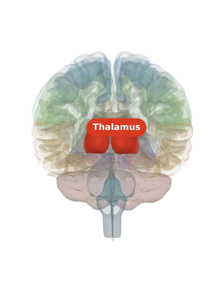
Not a separate shape in this atlas; lit: the part that holds it. Lit: the whole thalamus, on both sides. The reticular nucleus is only a sheet thick, laid over the outer surface of the thalamus; BodyParts3D has no shape for it (its only thalamic nuclei are the two geniculate bodies), so the whole thalamus is lit and nothing is drawn at a guessed point. Look at the lit thalamus’s outer face, the side turned towards the internal capsule. [1][2]
Lit here: the thalamus, the part that holds the reticular nucleus of the thalamus — see it on its own page.
It forms a capsule round the thalamus on its lateral side and is separated from the rest of the thalamus by the external medullary lamina, the thin layer of white matter that covers the thalamus’s lateral surface. [1][3]
Described in the rat, it lies between the external medullary lamina and the internal capsule, a sheet of GABA cells covering most of the lateral, dorsolateral and front (anterior) faces of the thalamus. [2]
It is counted with the zona incerta in the ventral thalamus (prethalamus), not with the dorsal thalamus whose relay nuclei it surrounds. [1][3]
It is laid out in sectors that face the nuclei they serve: a front (rostral) motor sector that also takes in limbic and prefrontal parts, a ventrocentral touch (somatosensory) sector, a ventrocaudal hearing sector and a dorsocaudal visual sector. [4]
What it does
All its cells use GABA. They receive glutamate from side branches of the fibres running between the thalamus and the cortex (thalamocortical fibres going out, corticothalamic fibres from cortical layer 6 coming in) and send inhibition back onto the thalamic relay neurons. [1][4]
It never projects to the cerebral cortex; it modulates what the other thalamic nuclei pass on, which is why it is described as gating thalamocortical transmission. [1][4]
It has long been thought to take part in attention; its subnetworks are proposed to carry bottom-up, top-down and between-nucleus attention effects. [4]
Rhythmic burst firing of its neurons, with the relay neurons they inhibit, generates sleep rhythms, among them the sleep spindles: short 11–16 Hz bursts that mark stage 2 non-REM sleep on an EEG. [4][5]
Connections
Inputs: the cerebral cortex and the dorsal thalamic nuclei. Outputs: back to the dorsal thalamic nuclei only. [1]
Other names
Abbreviated TRN, RTN, NRT or RT. It is not the reticular formation, the network of the brainstem’s core with a similar name. [1][6]
Size
The model has no shape for the reticular nucleus of the thalamus itself, so it is not measured here. The left thalamus that holds it measures about 37 mm front to back, 24 mm top to bottom and 24 mm side to side on this model (the box it fits in, not its volume). [7]
Facts describe anatomy and normal function only. The 3D model is the adult male BodyParts3D brain (credits and licence); real brains vary in size and shape, so its measurements describe one model, not anyone’s brain.