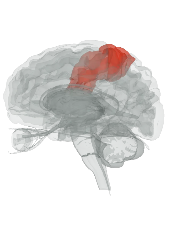
The paracentral lobule is at the top of the inner (medial) face of each hemisphere, where the precentral and postcentral gyri continue over the upper edge of the brain. Its front part belongs to the frontal lobe and its back part to the parietal lobe. [1]
Opens cut down the midline and seen from the left, so you look at the inner face of the right hemisphere: the lit precentral and postcentral gyri climb the outer face and fold over the top edge onto this inner face; the paracentral lobule is that folded-over stretch at the top, above the cingulate gyrus and in front of the precuneus. Drag to turn, pinch or scroll to zoom, tap any part to name it; .
Lit: the whole precentral and postcentral gyri, on both sides. The area is part of these gyri; its exact border is not drawn. It is the continuation of the precentral and postcentral gyri on the medial surface. BodyParts3D has no paracentral lobule shape, but its precentral and postcentral gyrus shapes run over onto that inner face (337 and 165 of their vertices lie within 5 mm of the midline), so both whole gyri are lit. [1][2]
It is the continuation of the precentral and postcentral gyri onto the medial surface of the cerebral hemisphere. [1]
It is framed by the cingulate sulcus: in front by the paracentral sulcus, a branch of the cingulate sulcus, and behind by the marginal sulcus. [1][3]
Where the central sulcus reaches the medial surface, it divides the lobule into a front part and a back part. On the medial face that groove is often not visible, so the border between the two lobes here is then judged from the cell layers (cytoarchitecture). [1]
The front part belongs to the frontal lobe and holds a little of Brodmann area 6, the supplementary motor area, because the paracentral sulcus does not line up with the precentral sulcus on the medial face. The back part belongs to the parietal lobe. [1]
Behind it, past the marginal branch of the cingulate sulcus, lies the precuneus. [4]
What it does
Its neurons serve the movement (motor) and sensation (sensory) innervation of the lower limb on the opposite side of the body. [1]
The back, parietal part deals with body sensation (somatosensation) of the far ends of the limbs. [1]
It also takes part in regulating body functions: control of urination and defecation, and blushing, which a 2024 study traced through the rise in cheek temperature when people watch themselves. [1][5]
The front, sensorimotor subdivision of the precuneus is wired to the paracentral lobule, the supplementary motor area, the premotor cortex and the insula. [4]
The model has no shape for the paracentral lobule itself, so it is not measured here. The whole left precentral and postcentral gyri that hold it measure, together, about 81 mm front to back, 66 mm top to bottom and 67 mm side to side on this model (the box they fit in, not their volume). [2]
Facts describe anatomy and normal function only. The 3D model is the adult male BodyParts3D brain (credits and licence); real brains vary in size and shape, so its measurements describe one model, not anyone’s brain.