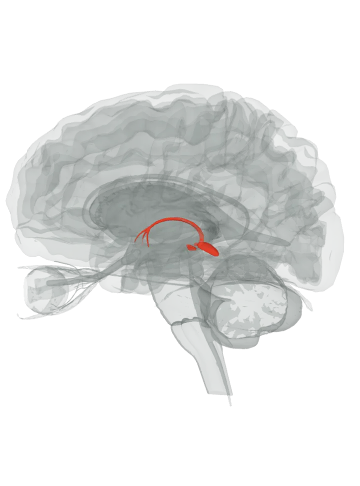
The epithalamus is the back (dorsal) part of the diencephalon, at the top back of the third ventricle, just above the midbrain. Its parts this model draws, the habenula, pineal gland, stria medullaris and posterior commissure, are lit together. [1][2]
All 4 are lit together, on both sides where they are paired; tap a name in the list to focus one. Drag to turn, pinch or scroll to zoom, tap any part to name it; .
Not lit: the habenular commissure, which joins the left and right habenula, and the anterior and posterior paraventricular nuclei of the thalamus (not the hypothalamus’s paraventricular nucleus, which has its own page). Both are named as epithalamus in the cited source, but neither is a separate shape in this 3D model, so no marker is placed at a guessed position. [1][2][3]
The cited source lists its parts as the habenular nuclei, the stria medullaris, the anterior and posterior paraventricular nuclei, the posterior commissure and the pineal gland; a shorter description in the same source names the habenular trigone, the pineal gland and the habenular commissure. [1]
The posterior commissure sits on the back border of the third ventricle, just below the pineal gland, near the corner where the ventricle narrows into the cerebral aqueduct. [5][6]
The stria medullaris runs back along the roof of the third ventricle, arching over the thalamus, then descends to end in the habenula; with the habenula and the habenular commissure it forms the habenular trigone. [7]
What it does
Its pineal gland makes and secretes melatonin; light reduces melatonin production and darkness increases it, which helps set sleep patterns. [8]
The habenula takes in signals mainly through the stria medullaris; its lateral part fires for unpleasant events and when an expected reward does not come, and in turn holds back the dopamine neurons of the substantia nigra and ventral tegmental area. [2][7]
Fibres of the pupillary light reflex cross in the posterior commissure to reach the Edinger–Westphal nuclei of both sides, which is why light in one eye narrows both pupils. [6][9]
Other names
The posterior commissure is also called the epithalamic commissure. [6]
Size
On this model the epithalamus together fit in a box about 44 mm front to back, 21 mm top to bottom and 15 mm side to side (the box they fit in, not their volume). [3]
Facts describe anatomy and normal function only. The 3D model is the adult male BodyParts3D brain (credits and licence); real brains vary in size and shape, so its measurements describe one model, not anyone’s brain.