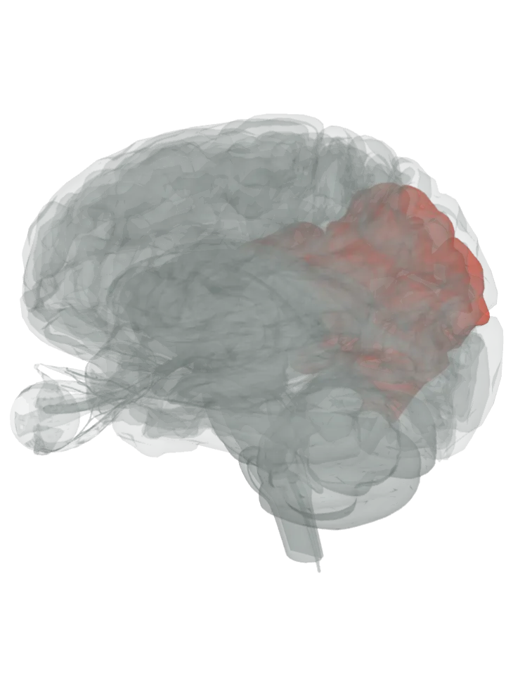
The inferior parietal lobule is the lower part of each parietal lobe, below the horizontal part of the intraparietal sulcus and behind the lower end of the postcentral sulcus. It is made of two gyri, the supramarginal gyrus in front and the angular gyrus behind, which are lit together. [1]
All 2 are lit together, on both sides where they are paired; tap a name in the list to focus one. Drag to turn, pinch or scroll to zoom, tap any part to name it; .
Not lit: the intraparietal sulcus, the groove that forms its upper border. A groove has no shape of its own in this 3D model, so it is not drawn here; its own page shows it by the parts on either side of it. [1][2][3]
Its supramarginal gyrus curves over the upturned back end of the lateral (Sylvian) fissure, and its angular gyrus over the back end of the superior temporal sulcus, so the lobule sits just above the back of the temporal lobe. [1]
Paired: there is one inferior parietal lobule in each hemisphere; this model lights both, each as its two gyri. [1][3]
What it does
It is concerned with language, mathematical operations and body image, the supramarginal and angular gyri in particular. [1]
It has also been involved in perceiving emotions in faces and in interpreting sensory information. [1]
Its angular gyrus is one of the hubs of the default mode network, the regions most active when a person is awake but not attending to the outside world. [5][6]
Other names
Also called Geschwind’s territory, after the American neurologist Norman Geschwind, and the subparietal district. Its two gyri correspond to Brodmann areas 40 (supramarginal) and 39 (angular). [1]
Size
On this model the inferior parietal lobule together fit in a box about 74 mm front to back, 64 mm top to bottom and 139 mm side to side (the box they fit in, not their volume). [3]
Facts describe anatomy and normal function only. The 3D model is the adult male BodyParts3D brain (credits and licence); real brains vary in size and shape, so its measurements describe one model, not anyone’s brain.