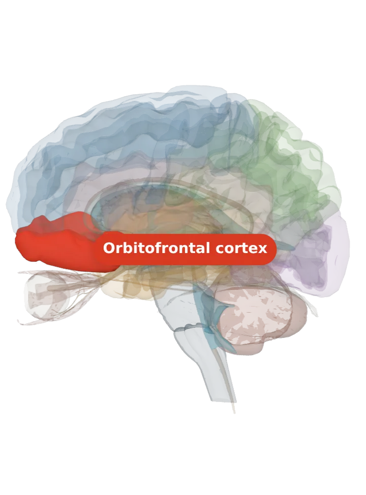
The gyrus rectus (straight gyrus) is the strip of the underside of each frontal lobe right beside the longitudinal fissure, on the inner side of the medial orbital gyrus and the olfactory tract. On the inner face of the hemisphere it continues into the superior frontal gyrus. [1]
Opens cut down the midline and seen from the left, so you look at the inner face of the right frontal lobe: the lit orbitofrontal cortex is the underside of the lobe, above the eye socket; the gyrus rectus is its strip right along the midline, the edge nearest you at the bottom front. Drag to turn, pinch or scroll to zoom, tap any part to name it; .
Lit: the whole orbitofrontal cortex, on both sides. The area is part of this cortex; its exact border is not drawn. It is the innermost of the gyri on the orbital surface, next to the midline. BodyParts3D has no gyrus rectus shape: it draws the orbital gyri of each side as one shape (lit here as the orbitofrontal cortex), which reaches the midline (553 of its vertices lie within 5 mm of it), so that whole shape is lit and the superior frontal gyrus is not. [2][3]
It is the portion of the lower frontal lobe immediately next to the longitudinal fissure, medial to the medial orbital gyrus and the olfactory tract. [1]
From the midline outward, the orbital surface reads: the gyrus rectus, then across the olfactory sulcus the medial orbital gyrus, then the anterior, posterior and lateral orbital gyri. [2]
The olfactory bulb and its stalk lie in the olfactory sulcus, which runs close to and parallel with the midline, along the outer side of the gyrus rectus. [4][1]
Brodmann area 11 covers most of the orbital gyri, the gyrus rectus and the most forward part of the superior frontal gyrus; it is the part of the orbitofrontal cortex over the medial half of the frontal lobe’s underside. [5]
What it does
Its encyclopaedia article says plainly that a specific function for the straight gyrus has not yet been brought to light. [1]
Brodmann area 11, which takes in the gyrus rectus, is involved in decision making, processing rewards and encoding new information into long-term memory. [5]
Connections
In the orbitofrontal cortex it belongs to, the back (caudal) part receives direct input from the piriform (smell) cortex and is the part most heavily connected with the amygdala. [2]
The model has no shape for the gyrus rectus itself, so it is not measured here. The whole left orbitofrontal cortex that holds it measures about 67 mm front to back, 30 mm top to bottom and 40 mm side to side on this model (the box it fits in, not its volume). [3]
Facts describe anatomy and normal function only. The 3D model is the adult male BodyParts3D brain (credits and licence); real brains vary in size and shape, so its measurements describe one model, not anyone’s brain.