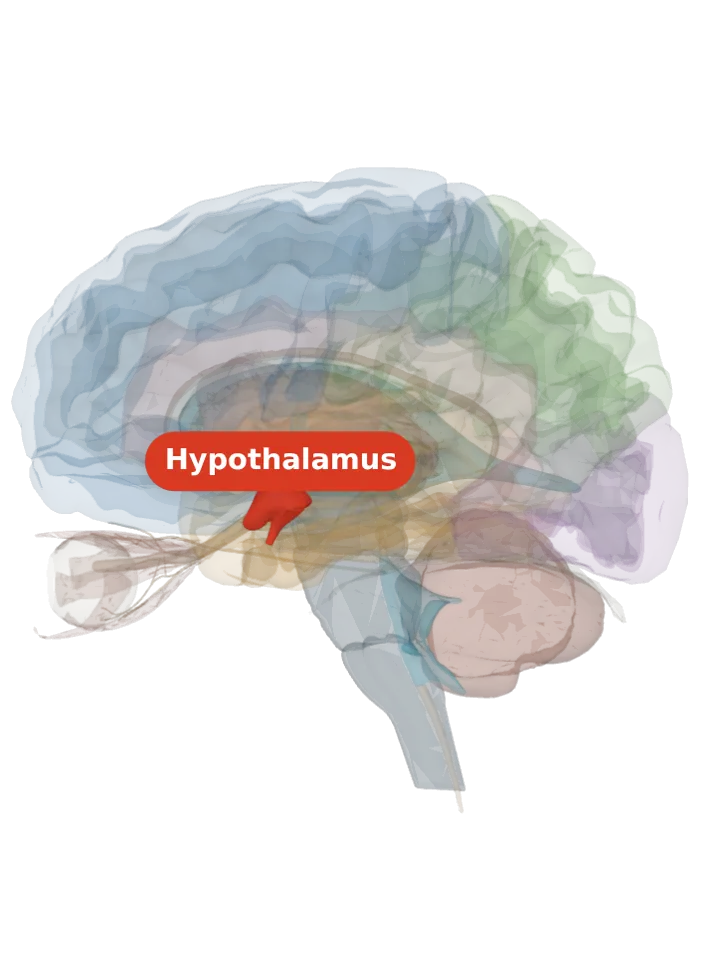
Where it is
- Of the hypothalamus’s three regions front to back (preoptic/anterior, tuberal, mammillary/posterior), it belongs to the middle, tuberal region, in its medial area, together with the dorsomedial and arcuate nuclei. [1]
- Its name gives its place: ventro-, the lower side, and medial, towards the midline; the dorsomedial nucleus lies above it, and the two border each other. [1][4]
- The arcuate nucleus lies against its lower outer (ventrolateral) edge; glucose-sensing neurons are packed into the narrow cell-poor strip between the two. [2]
- It has four subdivisions, anterior (VMHa), dorsomedial (VMHdm), ventrolateral (VMHvl) and central (VMHc), which differ in anatomy, chemistry and behaviour. [3]
What it does
- It is most commonly associated with satiety, the recognition of the feeling of fullness, and Wikipedia describes it as involved in ending hunger, fear, thermoregulation and sexual activity. [3]
- It holds glucose-sensing neurons, some excited and some inhibited by glucose, that respond to the body’s fat signals insulin and leptin and play an important role in the hormone and nerve responses that bring blood glucose back up when it falls too low. [2]
- Its ventrolateral part is packed with estrogen and progesterone receptors (co-expressed in nearly all its cells there); it plays a role in female mating posture (lordosis) and in estrogen-driven movement and heat production. [5][3]
- In mice, switching on estrogen-receptor neurons of the ventrolateral part sets off an immediate attack and silencing them stops naturally occurring attack, so it is also a hub for defensive and territorial behaviour. [5]
- Honest limit: the “satiety centre” label comes from rat lesion experiments. One researcher, Gold, reported that a precise cut confined to the nucleus did not change eating, which suggested passing fibres were behind the effect; other anatomical studies did not reproduce his result, and the label has held. [3]
Connections
- The stria terminalis carries fibres from the amygdala to the hypothalamus, including to its ventromedial nucleus. [1]
- It sends one of the strongest hypothalamic projections to the periaqueductal grey of the midbrain: of the Allen mouse brain atlas tracer injections ranked by the size of their endings in the periaqueductal grey, 6 of the top 10 were in the VMH. [5]
Other names
- Also called the ventromedial nucleus (VMN) or the ventromedial hypothalamic nucleus; VMN, VMH and ventromedial hypothalamus are used interchangeably. It is not the ventromedial nucleus of the spinal cord, nor the ventromedial prefrontal cortex. [3]
Size
- The model has no shape for the ventromedial hypothalamus itself, so it is not measured here. The hypothalamus that holds it measures about 18 mm front to back, 20 mm top to bottom and 17 mm side to side on this model (the box it fits in, not its volume). [6]