The central amygdala (the central nucleus of the amygdala) is a small nucleus in the upper (dorsal) part of each amygdala, near where the stria terminalis leaves it. It is the amygdala’s main output station, passing its alarm on to the hypothalamus and the brainstem. [1][2]
Opens cut by a front (coronal) slice through the amygdala, which holds the central amygdala. Drag to turn, pinch or scroll to zoom, tap any part to name it; .
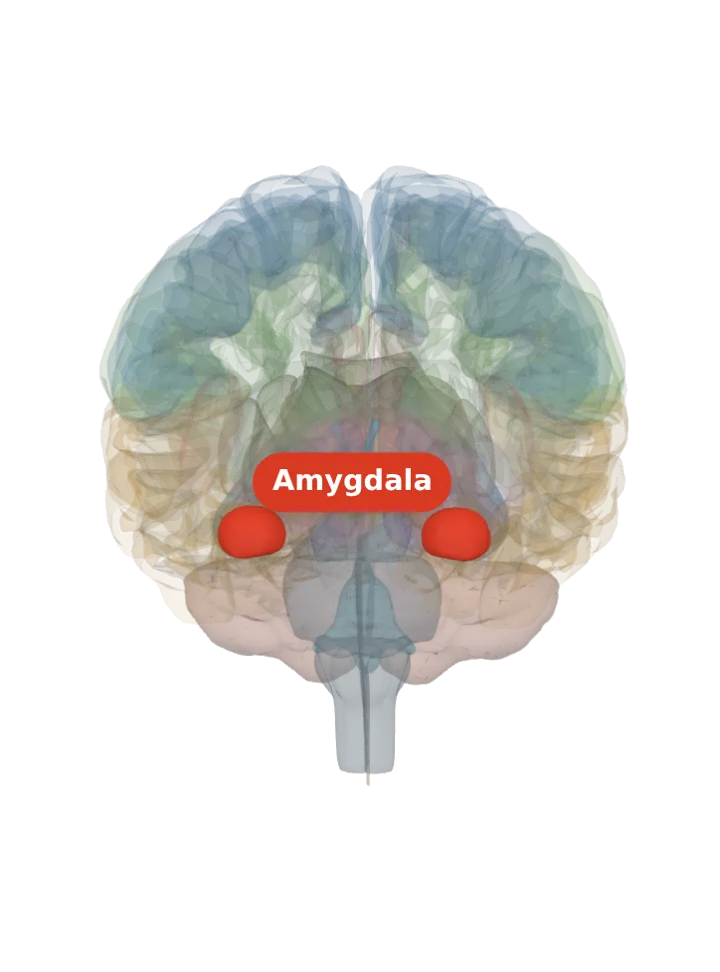
Not a separate shape in this atlas; lit: the part that holds it. Lit: the whole amygdala, on both sides. The central nucleus is a small nucleus inside the amygdala, about 3% of its volume; BodyParts3D draws each amygdala as one shape with no nuclei in it, so the whole amygdala is lit and nothing is drawn at a guessed point. Look at the upper (dorsal) side of the lit amygdala, the side nearest the stria terminalis. [1][3]
Lit here: the amygdala, the part that holds the central amygdala — see it on its own page.
It lies in the dorsal amygdala, next to the cortical and medial nuclei and near the stria terminalis, a major output pathway of the amygdala. [1]
With the cortical and medial nuclei and the anterior amygdaloid area it forms the centromedial nuclear group, which turns sensory information into behavioural and autonomic responses. [1]
Inhibitory intercalated cells lie between it and the basolateral nuclei; medial clusters of them sit towards the central amygdala. [4]
What it does
It is the major output nucleus of the amygdala, and it also receives and processes pain information. [2]
The central nuclei set off many fear responses: defensive behaviour (freezing or escape), autonomic changes in blood pressure and heart rate, and stress-hormone release. [5]
It produces the body’s side of emotion mainly through outputs to the lateral hypothalamus and the brainstem, and takes part in conscious perception of emotion through the ventral amygdalofugal pathway to the anterior cingulate, orbitofrontal and prefrontal cortex. [2]
In the stress response, signals passed from the lateral amygdala to the central nucleus reach the hypothalamus; for a threat that is only expected, amygdala GABA neurons start the response by silencing cells that normally hold the paraventricular nucleus back. [6]
Honest limit: most of this circuit work was done in rats and other animals; in rats, for example, the central nucleus, but not the basolateral regions, is needed for some appetitive (reward) learning. [5]
Connections
In: its main input is the basolateral complex, which sends it what it has processed; some sensory neurons also end in it directly. [7][5]
In a human MRI atlas it is the smallest of the nine amygdala subregions measured: 47–53 mm³, about 3% of an amygdala of 1,582–1,804 mm³. [3]
The model has no shape for the central amygdala itself, so it is not measured here. The left amygdala that holds it measures about 18 mm front to back, 13 mm top to bottom and 17 mm side to side on this model (the box it fits in, not its volume). [9]
Facts describe anatomy and normal function only. The 3D model is the adult male BodyParts3D brain (credits and licence); real brains vary in size and shape, so its measurements describe one model, not anyone’s brain.