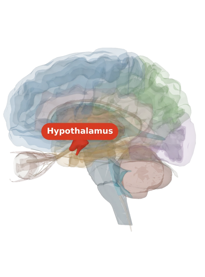
The dorsomedial hypothalamus is a nucleus high in the middle (tuberal) region of the hypothalamus, near the midline and directly above the ventromedial nucleus. It relays the body clock’s daily timing to sleep, feeding and body temperature. [1][2][3]
Opens cut by a side (sagittal) slice through the hypothalamus, which holds the dorsomedial hypothalamus. Drag to turn, pinch or scroll to zoom, tap any part to name it; .
Not a separate shape in this atlas; lit: the part that holds it. Lit: the whole hypothalamus. The dorsomedial hypothalamus is a nucleus inside the hypothalamus, not a separate organ; BodyParts3D has no shape for it, so the whole hypothalamus is lit and nothing is drawn at a guessed point. Look at the upper half of the middle of the lit hypothalamus, near the midline, above the ventromedial nucleus. [3][1]
Lit here: the hypothalamus, the part that holds the dorsomedial hypothalamus — see it on its own page.
It belongs to the middle, tuberal region of the hypothalamus, in its medial area, together with the ventromedial and arcuate nuclei. [1]
Its name gives its place: dorso-, the upper side, and medial, towards the midline; it sits above the ventromedial nucleus, and the two border each other. [1][2]
In rat brain sections it is found by the landmarks around it: the third ventricle, the fornix, the mammillothalamic tract and the median eminence. [2]
What it does
It is involved in feeding, drinking, body-weight regulation and circadian activity, and is a necessary link for many behavioural and physiological daily rhythms to show. [3]
When food is offered only at a fixed time of day, animals start to wake and move about before it arrives; in rats, lesions of the DMH weakened this food entrainment of wakefulness, movement and core body temperature, and it may hold its own food-entrainable oscillator (not yet shown in a dish). [3]
It is also known as a stress centre: in rats, quieting it with the drug muscimol blocked 85% of the heart-rate rise and 68% of the blood-pressure rise to a puff of air. [3]
Stress raises body temperature through a path from the DMH to premotor neurons in the rostral raphe pallidus of the medulla, which drive heat production. [3][4]
Honest limit: the evidence is from rats and mice, and later lesion work found that the DMH cannot be the only place a food-timed clock lives, since food anticipation can survive without it. [5][3]
Connections
It receives the body clock’s signal from the suprachiasmatic nucleus, both directly and by way of the subparaventricular zone. [3]
It sends information on to the ventrolateral preoptic area, the sleep-promoting group at the front of the hypothalamus. [3]
Other names
Also called the dorsomedial hypothalamic nucleus, short form DMH; Latin nucleus dorsomedialis areae hypothalamicae intermediae. It is not the dorsomedial (mediodorsal) nucleus of the thalamus. [3]
Size
The model has no shape for the dorsomedial hypothalamus itself, so it is not measured here. The hypothalamus that holds it measures about 18 mm front to back, 20 mm top to bottom and 17 mm side to side on this model (the box it fits in, not its volume). [6]
Facts describe anatomy and normal function only. The 3D model is the adult male BodyParts3D brain (credits and licence); real brains vary in size and shape, so its measurements describe one model, not anyone’s brain.