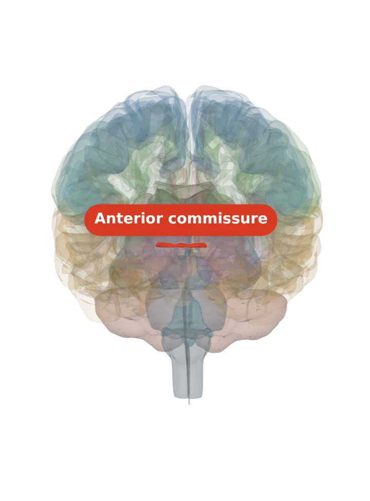
The bed nucleus of the stria terminalis (BNST) is a small group of nuclei in the basal forebrain, one on each side, far from the amygdala itself: it sits on the anterior commissure where that crosses the midline, under the caudate nucleus, with the internal capsule on its outer side. About 74 mm³ each, it is a relay in the stress response. [1][2]
Opens cut by a front (coronal) slice through the anterior commissure, which holds the bed nucleus of the stria terminalis. Drag to turn, pinch or scroll to zoom, tap any part to name it; .
Not a separate shape in this atlas; lit: the part that holds it. Lit: the whole anterior commissure. The bed nucleus lies against the lit anterior commissure: it sits on top of the commissure at the level where it crosses the midline, one on each side, under the caudate nucleus and with the stria terminalis along its inner side. BodyParts3D has no shape for the bed nucleus, so the commissure is lit and nothing is drawn at a guessed point; look just above each half of the lit bar. [1]
Lit here: the anterior commissure, the part the bed nucleus of the stria terminalis lies against — see it on its own page.
It lies at the level where the anterior commissure crosses, in the basal forebrain: the caudate nucleus is its upper border at most levels, the internal capsule its outer border at all levels, and the anterior commissure lies below its middle stretch. [1]
It runs from the head of the caudate nucleus at the front to the reticular nucleus of the thalamus at the back; it is compact at the front, triangular at the anterior commissure and elongated behind it. [1]
Wikipedia’s short version: a collection of nuclei at the base of the lateral ventricles, in front of the septum. [2]
What it does
It is thought to act as a relay in the hypothalamic–pituitary–adrenal (HPA) axis, adjusting that axis’s activity in response to acute stress. [2]
Its part in the stress response is tied to time: a sudden fright lasting under ten minutes does not activate it. [2]
Honest limit: much of the detailed work on it, such as its part in controlling feeding, was done in rodents, and in living people its small size and low contrast make it hard or impossible to pick out on routine MRI. [2][1]
Connections
The stria terminalis, the amygdala’s major output pathway, runs along it, carrying fibres from the amygdala to the septal nuclei, hypothalamus and thalamus and back. [2]
With the central amygdala it forms the extended amygdala, a grouping made by shared connections and chemical staining whose borders are not sharp under the microscope; one cited source adds the shell of the nucleus accumbens. [4]
The bed nucleus, the hypothalamic nuclei and the septal nuclei sit close together and share many projections. [2]
Inside it
Mapped across ten human brains, it has four subdivisions: a central, a dorsal, a medial and a posterior part. [1]
Other names
It was first described in 1923 by Johnston as a “stria bed”, then counted as part of the caudate nucleus; its links with the amygdala later gave rise to the idea of an extended amygdala. Short forms: BNST or BST. [1]
Size
Mean volume about 74 mm³ in each hemisphere (shrinkage-corrected, ten post-mortem brains), with subdivisions from about 8 mm³ (central) to 31 mm³ (dorsal); that study found no difference between the sexes or the hemispheres. [1]
The model has no shape for the bed nucleus of the stria terminalis itself, so it is not measured here. The anterior commissure that holds it measures about 5 mm front to back, 3 mm top to bottom and 24 mm side to side on this model (the box it fits in, not its volume). [5]
Facts describe anatomy and normal function only. The 3D model is the adult male BodyParts3D brain (credits and licence); real brains vary in size and shape, so its measurements describe one model, not anyone’s brain.