The corona radiata is a sheet of white matter in each cerebral hemisphere, above the basal ganglia: going down it continues as the internal capsule, going up as the centrum semiovale under the cortex. Its ascending and descending axons carry most of the traffic to and from the cerebral cortex. [1]
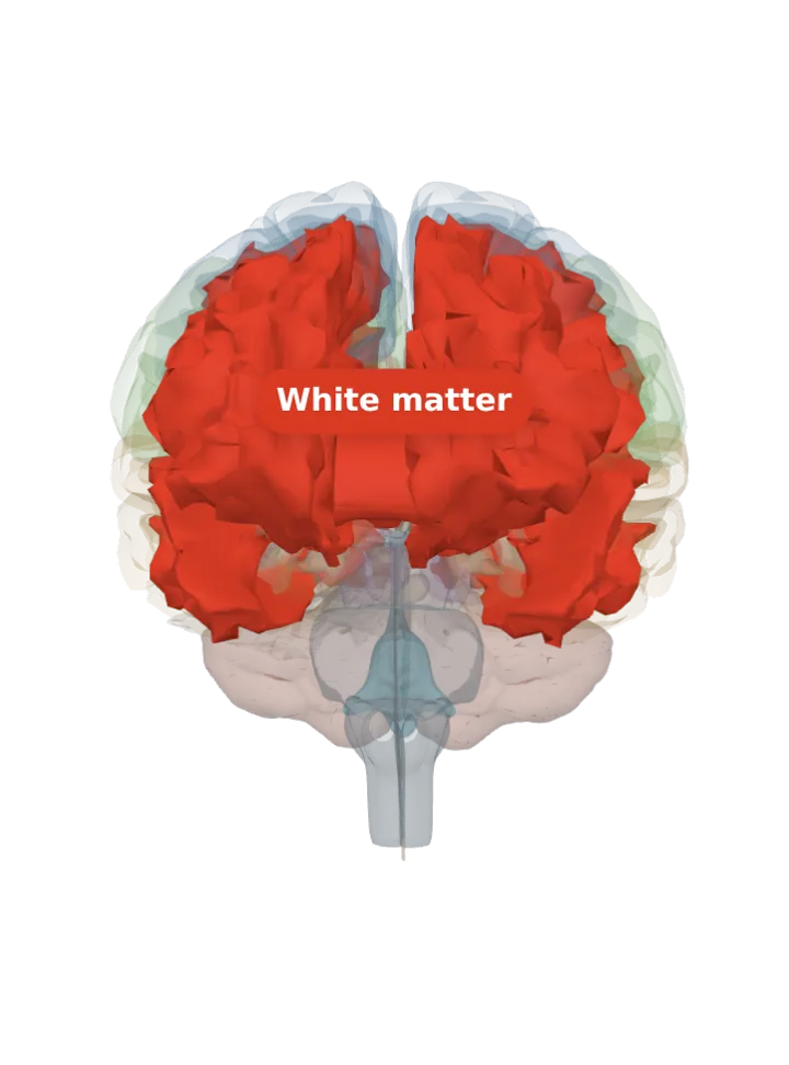
Opens cut by a front (coronal) slice through the white matter, which holds the corona radiata. Drag to turn, pinch or scroll to zoom, tap any part to name it; .
Not a separate shape in this atlas; lit: the part that holds it. Lit: the whole white matter. The corona radiata is part of the lit white matter: the stretch of it just above the internal capsule, where the capsule’s fibres fan out towards the cortex. The BodyParts3D model draws each hemisphere’s white matter as one shape with no corona radiata of its own, so the white matter that holds it is lit and nothing is drawn at a guessed border. [1][2][3]
Lit here: the white matter, the part that holds the corona radiata — see it on its own page.
Where it is
It fans out from the internal capsule: above the basal ganglia the internal capsule’s fibres spread out as the corona radiata, and below the basal ganglia they gather into the crus of the cerebral peduncle in the midbrain. [2]
Going down it continues as the internal capsule; going up it continues as the centrum semiovale, the central white core under the cortex of each hemisphere. [1][4]
Projection fibres radiate out from the cortex through it and come together lower down; the same fibres pass between it and the brainstem through the internal capsule. [1]
What it does
Its sheet of ascending and descending axons carries most of the neural traffic to and from the cerebral cortex. [1]
Three projection tracts run in it: the corticospinal tract (motor commands to the spinal cord), the corticobulbar tract (to the brainstem nuclei of the cranial nerves) and the corticopontine tract (to the pons). [1][5]
Projection fibres in it are both afferent, bringing information to the cortex, and efferent, carrying it away. [1]
Connections
The corticofugal projection, the fibres leaving the cortex for deeper parts, runs through the corona radiata and then through the internal capsule. [1]
Inside it
Mostly myelinated axons, which give white matter its pale colour; it holds no nuclei of its own. [6][1]
Size
The model has no shape for the corona radiata itself, so it is not measured here. The white matter that holds it measures about 163 mm front to back, 100 mm top to bottom and 125 mm side to side on this model (the box it fits in, not its volume). [3]
Facts describe anatomy and normal function only. The 3D model is the adult male BodyParts3D brain (credits and licence); real brains vary in size and shape, so its measurements describe one model, not anyone’s brain.