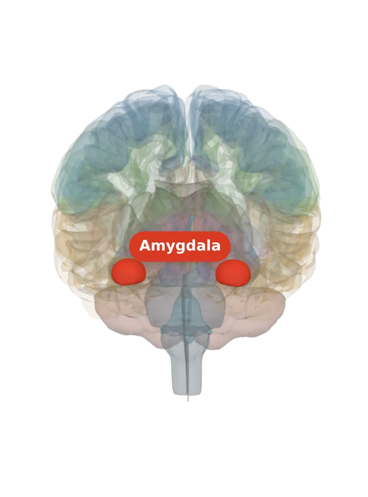
The basolateral amygdala is the deep, larger nucleus group of each amygdala, in the inner (medial) part of the temporal lobe just in front of the hippocampus: the lateral, basal and accessory basal nuclei together. It is the amygdala’s input side, where sensory signals arrive and are linked to what followed them. [1][2][3]
Opens cut by a front (coronal) slice through the amygdala, which holds the basolateral amygdala. Drag to turn, pinch or scroll to zoom, tap any part to name it; .
Not a separate shape in this atlas; lit: the part that holds it. Lit: the whole amygdala, on both sides. The basolateral amygdala is a group of nuclei inside the amygdala, not a separate organ; BodyParts3D draws each amygdala as one shape with no nuclei in it, so the whole amygdala is lit and nothing is drawn at a guessed point. Look at its deep core: in a human MRI atlas the lateral nucleus alone is about a fifth of the amygdala. [1][4]
Lit here: the amygdala, the part that holds the basolateral amygdala — see it on its own page.
It consists of the lateral, basal and accessory basal nuclei of the amygdala, also called the basolateral complex or basolateral nuclear complex. [1][3]
It is the amygdala’s deep nucleus group; the central, medial and cortical nuclei form a superficial group around it. [2]
A net of small inhibitory cells, the intercalated cells, wraps it and lies between it and the central nucleus. [5][3]
The amygdala itself sits medially in each temporal lobe, just in front of the hippocampus, so there is one basolateral amygdala on each side. [3]
What it does
Its lateral nucleus receives most of the amygdala’s sensory information, which arrives directly from temporal-lobe structures including the hippocampus and the primary auditory cortex. [1]
In fear conditioning, sensory signals reach the basolateral complex, particularly the lateral nucleus, where they are associated with memories of what followed; its neurons are responsible for forming the conditioned fear memory. [3][6]
The amygdala, especially its basolateral nuclei, strengthens memory for events that were emotionally arousing, as shown by James McGaugh’s laboratory and others. [3]
It learns good associations as well as bad: in female mice it is the key centre for learning to like an odour that has been paired with an attractive pheromone. [3]
Honest limit: most of the circuit work here was done in rats and mice; in people, routine MRI has too little contrast to see the nuclei, so human studies such as the one measuring their sizes map them with a probabilistic atlas. [6][4]
Connections
It connects extensively with higher-order cortex in the prefrontal, temporal and insular lobes, and with the hippocampus. [3]
Its glutamate-releasing neurons project to the nucleus accumbens, both its shell and its core. [3]
It receives dense neuromodulatory input from the ventral tegmental area, the locus coeruleus and the basal forebrain, which matters for associative learning. [1]
Other names
Also called the basolateral complex or the basolateral nuclear complex; short form BLA. [1][6]
Size
In a human MRI atlas the lateral nucleus is about 21–22% of the amygdala’s volume (333–391 mm³), the dorsal and intermediate basal nucleus 12–13%, the ventral basal and paralaminar nuclei 7–8%, and the basomedial nucleus about 7%: together roughly half of an amygdala of 1,582–1,804 mm³. [4]
The model has no shape for the basolateral amygdala itself, so it is not measured here. The left amygdala that holds it measures about 18 mm front to back, 13 mm top to bottom and 17 mm side to side on this model (the box it fits in, not its volume). [8]
Facts describe anatomy and normal function only. The 3D model is the adult male BodyParts3D brain (credits and licence); real brains vary in size and shape, so its measurements describe one model, not anyone’s brain.