Where are the ventral lateral and ventral anterior nuclei?
The ventral anterior (VA) and ventral lateral (VL) nuclei sit in the lower (ventral) tier of the thalamus’s outer part, in front of the ventral posterior nucleus: VA at the front, VL just behind it. Together, often called the motor thalamus, they pass the output of the basal ganglia and the cerebellum on to the motor cortices. [1][2][3][4]
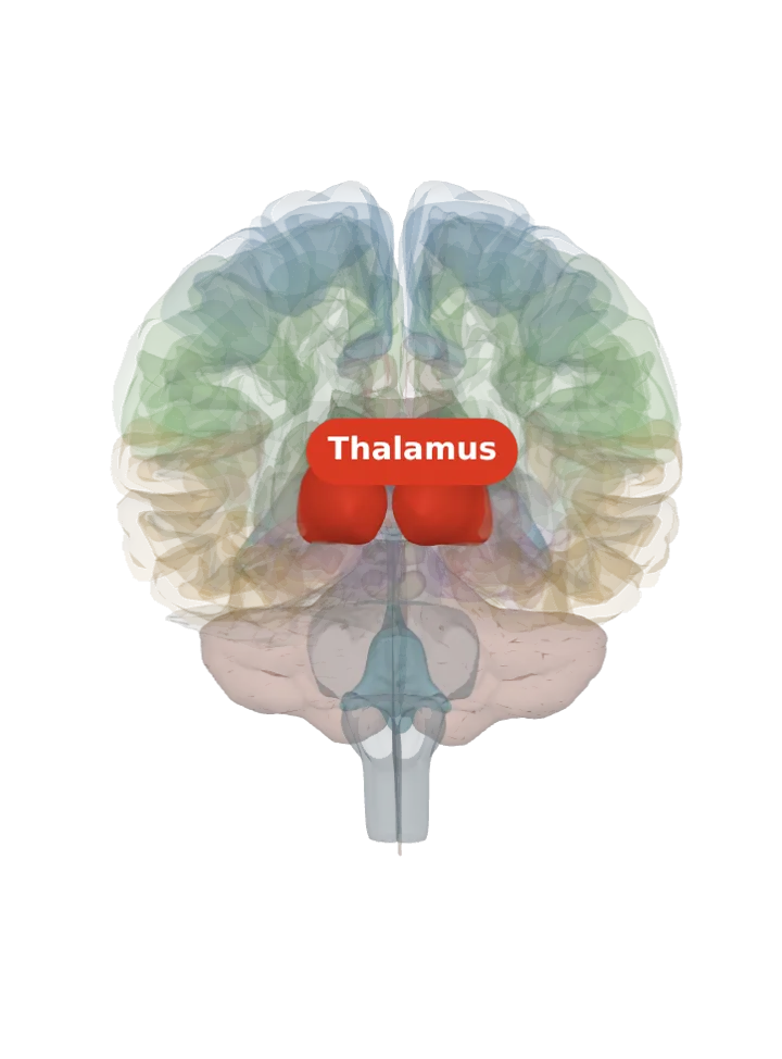
Opens cut by a front (coronal) slice through the thalamus, which holds the ventral lateral and ventral anterior nuclei. Drag to turn, pinch or scroll to zoom, tap any part to name it; .
Not a separate shape in this atlas; lit: the part that holds it. Lit: the whole thalamus, on both sides. The ventral anterior and ventral lateral nuclei are the front of the thalamus’s lower outer tier; BodyParts3D has no shape for it (its only thalamic nuclei are the two geniculate bodies), so the whole thalamus is lit and nothing is drawn at a guessed point. Look at the lower outer front of the lit thalamus. [1][2]
Lit here: the thalamus, the part that holds the ventral lateral and ventral anterior nuclei — see it on its own page.
Where it is
The Y-shaped internal medullary lamina divides each thalamus into anterior, medial and lateral groups; the ventral group is split into ventral anterior, ventral lateral and ventral posterior nuclei, front to back. [1]
The ventral lateral nucleus has an anterior part (VLa), which takes the pallidal input and projects mainly to the premotor cortex, and a posterior part (VLp), which takes the cerebellar input and is the main relay to the motor cortex; a ventral medial part is also named. [2][4]
What it does
The ventral anterior nucleus receives the basal ganglia’s output, its main input coming from the globus pallidus, and sends fibres to the premotor cortex for the starting and planning of movement. [3]
The ventral lateral nucleus receives the basal ganglia (the substantia nigra and the globus pallidus) and the cerebellum, through the dentatothalamic tract, and sends its output to the primary motor and premotor cortex; it helps coordinate, plan and learn movement. [2]
The two inputs mostly arrive in separate territories and with opposite signs: the basal ganglia’s GABA input (from the globus pallidus and the substantia nigra’s pars reticulata) inhibits the front territories, and the cerebellum’s glutamate input excites the back one (VLp). [4]
Here the basal ganglia loop closes: freed from the globus pallidus’s brake, the ventral anterior and ventral lateral nuclei excite the motor and premotor cortex, amplifying the movement the cortex has chosen. [2][3]
Connections
The territories that receive the basal ganglia are mainly connected with premotor and associative cortex, the cerebellar territory with the primary motor cortex; input from motor areas of the cortex reaches all of them. [4]
Short forms VA and VL (VLa and VLp for the two parts of VL); together with the ventral medial nucleus they are what studies call the motor thalamus. [2][3][4]
Size
The model has no shape for the ventral lateral and ventral anterior nuclei itself, so it is not measured here. The left thalamus that holds it measures about 37 mm front to back, 24 mm top to bottom and 24 mm side to side on this model (the box it fits in, not its volume). [5]
Facts describe anatomy and normal function only. The 3D model is the adult male BodyParts3D brain (credits and licence); real brains vary in size and shape, so its measurements describe one model, not anyone’s brain.