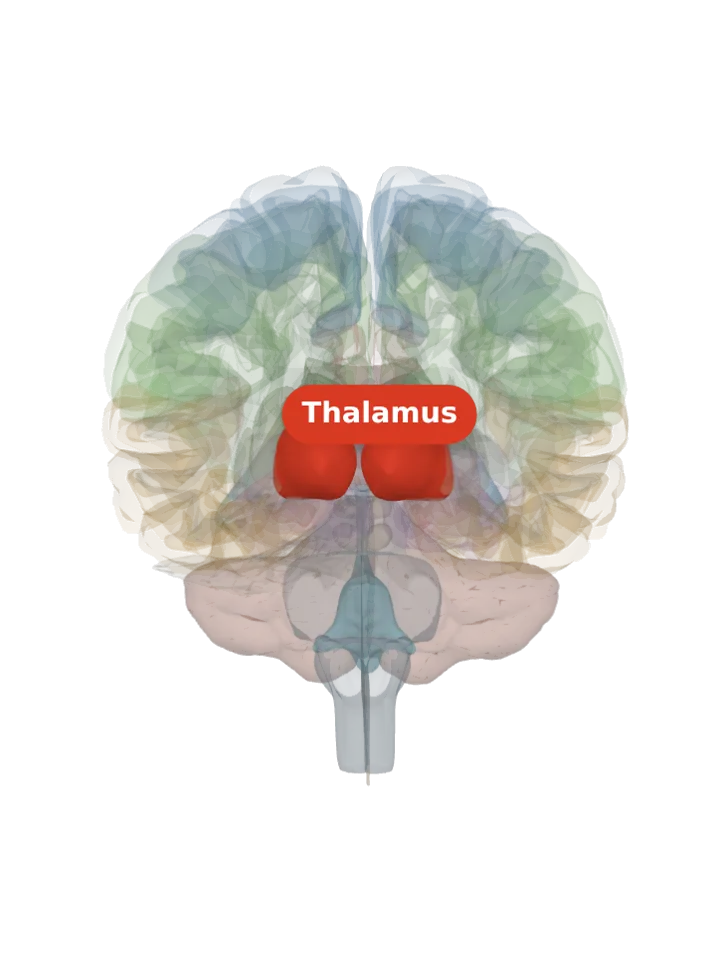
The intralaminar nuclei are cell groups embedded inside the Y-shaped internal medullary lamina, the thin sheet of white matter that splits each thalamus into its anterior, medial and lateral groups. The largest, the centromedian nucleus, lies in its back part, just below the mediodorsal nucleus. [1][2][3]
Opens cut by a front (coronal) slice through the thalamus, which holds the intralaminar nuclei. Drag to turn, pinch or scroll to zoom, tap any part to name it; .
Not a separate shape in this atlas; lit: the part that holds it. Lit: the whole thalamus, on both sides. The intralaminar nuclei are strung along the dividing sheet deep inside the thalamus; BodyParts3D has no shape for it (its only thalamic nuclei are the two geniculate bodies), so the whole thalamus is lit and nothing is drawn at a guessed point. Look at the inner core of the lit thalamus, between its middle and outer parts. [1][3]
Lit here: the thalamus, the part that holds the intralaminar nuclei — see it on its own page.
Where it is
They are usually split into two groups: a front (rostral) group, the central lateral, central medial and paracentral nuclei, and a back (caudal) group, the centromedian and parafascicular nuclei; some sources add a central dorsal nucleus. [1]
The centromedian nucleus sits in the medial thalamus, bordered above by the mediodorsal nucleus, on its inner side by the parafascicular nucleus and behind by the pulvinar; there is one on each side. [3]
What it does
The centromedian/parafascicular complex is involved in attention and general arousal, including control of the level of cortical activity; a 7-tesla fMRI study of healthy volunteers found it active in the preparation and expectancy phase of a task. [4][3]
They relay the brainstem’s arousal signals on to the cortex, and general anaesthetics specifically suppress their activity, the centromedian nucleus included. [3][5]
They are also thought to take part in the emotional and autonomic responses to pain: they receive the spinothalamic tract and the periaqueductal grey. [1]
Connections
Inputs come from the brainstem, the spinal cord and the cerebellum; the centromedian nucleus also receives the cerebral cortex, the vestibular nuclei, the globus pallidus, the superior colliculus and the reticular formation. [1][3]
Outputs: the centromedian nucleus sends fibres to the putamen and the subthalamic nucleus; the group as a whole projects to the hypothalamus, amygdala and limbic lobe, and its links with the cortex and the basal ganglia run both ways. [3][1]
Other names
Also called the intralaminar thalamic nuclei (ITN); the centromedian and parafascicular nuclei together are often written CM-Pf. [1][4]
Size
Each centromedian nucleus is under 10 mm in every dimension: about 310 cubic millimetres holding some 664,000 neurons, about 2,200 per cubic millimetre. [3]
The model has no shape for the intralaminar nuclei itself, so it is not measured here. The left thalamus that holds it measures about 37 mm front to back, 24 mm top to bottom and 24 mm side to side on this model (the box it fits in, not its volume). [6]
Facts describe anatomy and normal function only. The 3D model is the adult male BodyParts3D brain (credits and licence); real brains vary in size and shape, so its measurements describe one model, not anyone’s brain.