brainlocation

For learning, not medical advice.

All parts of the brain › Functions › Pain

What part of the brain processes pain?

Pain is processed in several places at once, not in one pain centre. [1][2][3][4]

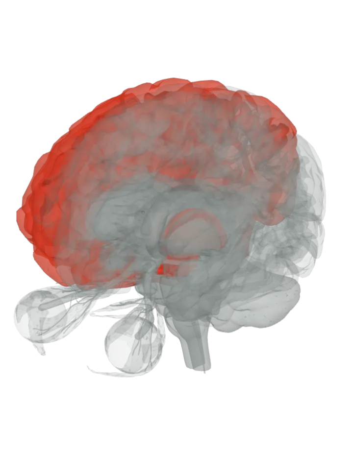
The thalamus, the postcentral gyrus, the insula, the cingulate gyrus, the midbrain and the prefrontal cortex lit in red on a 3D model of the brain, seen from the front left and above

Loading the 3D brain…

Lit: whole parts; region borders not drawn.

Slice

All 6 parts are lit together. Drag to turn, pinch or scroll to zoom, tap a part on the brain or a name in the list to focus it; .

Signals from pain receptors (nociceptors) climb the spinothalamic tract to the thalamus, which passes them on to the cortex: the primary and secondary somatosensory cortex on the postcentral gyrus and its operculum work out where the pain is and how strong, while the insula and the anterior cingulate cortex carry how unpleasant it feels. The periaqueductal grey in the midbrain is the main control centre for turning incoming pain down. [1][2][3][4]

The anterior cingulate cortex is a region on a lit gyrus, not a separate shape. The 3D model draws only whole gyri, so the gyrus that holds each area is lit; the area’s exact border is not drawn.

The secondary somatosensory cortex is a region on the postcentral gyrus, not a separate shape. The 3D model has no shape for it, so the whole postcentral gyrus is lit; the region’s exact border is not drawn.

How they work together

  • How it gets there: pain fibres enter the spinal cord, cross to the other side through the anterior white commissure and climb the spinothalamic tract to the thalamus, so the left half of the brain feels pain from the right side of the body. [1][12]
  • Pain is defined as an unpleasant sensory and emotional experience tied to actual or potential tissue damage, which is why it lights both sensory areas (the somatosensory cortex) and emotional ones (the insula and the anterior cingulate cortex). [1]
  • Two sides of one pain: in two PET studies by the same group, changing only how intense a heat felt moved activity in the primary somatosensory cortex, while changing only how unpleasant it felt moved the anterior cingulate cortex. [6][7]
  • A pattern, not a spot: across four studies of 114 people, a brain-scan pattern spanning the thalamus, insula, secondary somatosensory cortex, anterior cingulate cortex and periaqueductal grey told painful heat from mere warmth with 94% or better sensitivity and specificity in its first study. [4]
  • The brain turns pain down by itself: the periaqueductal grey signals the raphe nuclei, whose serotonin fibres run down to the dorsal horn of the spinal cord and damp pain signals there before they climb on. [3][2]
  • Honest limit: these are networks seen in imaging and animal work, not single centres; the scans give brief heat to small groups of healthy volunteers (ten in one PET study, 14 in another), the stimulation work on the periaqueductal grey was done in rats, and the ventral posterolateral nucleus, S2, the anterior cingulate cortex, the dorsolateral prefrontal cortex and the periaqueductal grey have no separate shapes on this model, so whole parts are lit, not exact borders. [6][9][8][13]

Pain shares the thalamus and postcentral gyrus with touch, but fine touch climbs its own route, the dorsal columns, while pain and temperature climb the spinothalamic tract; the touch page lights the whole body-feeling network. [14][12] What part of the brain feels touch, temperature and pain?

Itch travels the same road as pain, its unmyelinated fibres using the same nerve bundle and spinothalamic tract, but pain sets off a reflex to pull away and itch a scratch reflex. [15] What part of the brain feels an itch?

Sources

  1. Wikipedia: Pain
  2. Wikipedia: Nociception
  3. Wikipedia: Periaqueductal gray
  4. Wager et al. 2013, An fMRI-based neurologic signature of physical pain (N Engl J Med, free in PMC)
  5. Coghill et al. 1999, Pain intensity processing within the human brain: a bilateral, distributed mechanism (J Neurophysiol, PubMed abstract)
  6. Hofbauer et al. 2001, Cortical representation of the sensory dimension of pain (J Neurophysiol, PubMed abstract)
  7. Rainville et al. 1997, Pain affect encoded in human anterior cingulate but not somatosensory cortex (Science, PubMed abstract)
  8. Reynolds 1969, Surgery in the rat during electrical analgesia induced by focal brain stimulation (Science, PubMed abstract)
  9. Lorenz, Minoshima & Casey 2003, Keeping pain out of mind: the role of the dorsolateral prefrontal cortex in pain modulation (Brain, PubMed abstract)
  10. Wikipedia: Secondary somatosensory cortex
  11. Wikipedia: Operculum (brain)
  12. Wikipedia: Spinothalamic tract
  13. BodyParts3D (DBCLS), the atlas this 3D model is built from
  14. Wikipedia: Dorsal column–medial lemniscus pathway
  15. Wikipedia: Itch

Facts describe anatomy and normal function only. A part lit here takes part in feeling pain; it does not do it alone, and it does other jobs too. The 3D model is the adult male BodyParts3D brain (credits and licence); on it the brainstem includes the midbrain, pons and medulla, so lighting the brainstem lights all three.