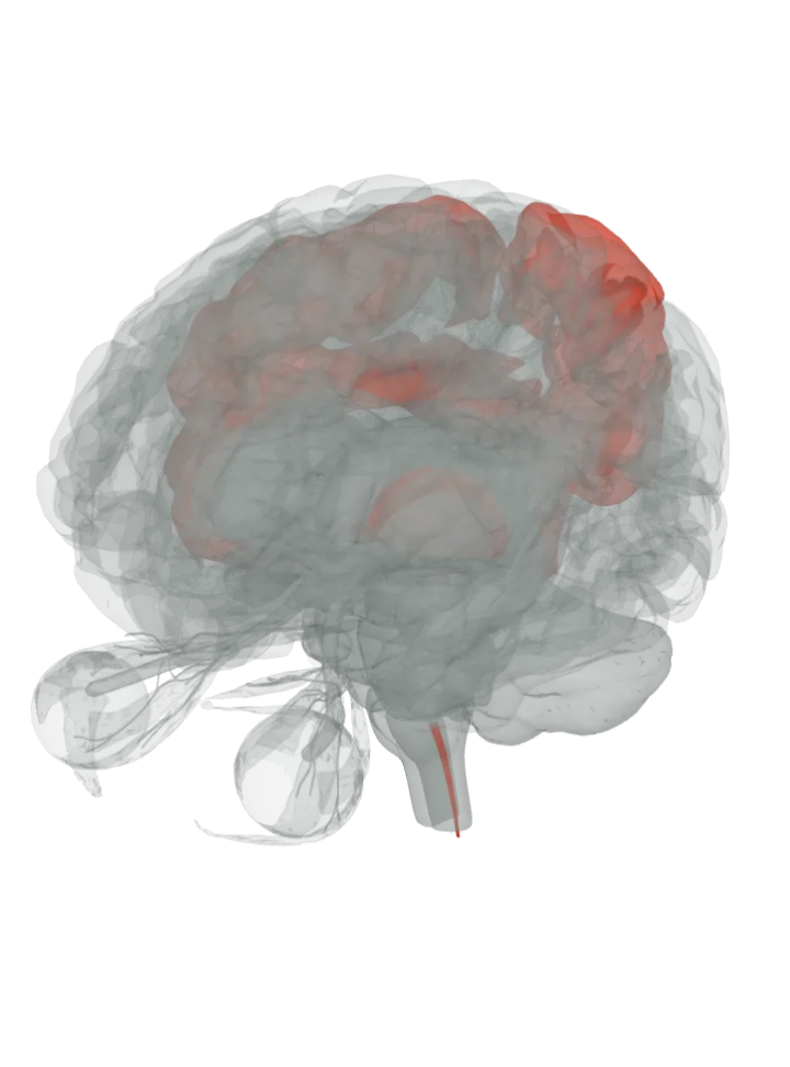
No single itch centre. Itch starts in thin unmyelinated nerve fibres in the skin, is passed on by a dedicated set of cells in the spinal cord’s dorsal horn and climbs the spinothalamic tract to the thalamus, the same route as pain. In the cortex it lights the somatosensory cortex on the postcentral gyrus, the insula and the anterior cingulate cortex, with motor areas that prepare the scratch. [1][2][3][4]
All 5 parts are lit together. Drag to turn, pinch or scroll to zoom, tap a part on the brain or a name in the list to focus it; .
The anterior cingulate cortex is a region on a lit gyrus, not a separate shape. The 3D model draws only whole gyri, so the gyrus that holds each area is lit; the area’s exact border is not drawn.
How they work together
Itch and pain share a road: their unmyelinated fibres both start in the skin and use the same nerve bundle and spinothalamic tract, but pain sets off a reflex to pull away and itch a scratch reflex. [1]
Separate lanes on the shared road: in primates, histamine and cowhage, a plant whose hairs itch without histamine, excite different groups of spinothalamic neurons that keep the two kinds of itch apart. [6]
Why scratching helps: in primates, spinothalamic neurons that fired to histamine fired less after the skin was scratched, an inhibition that seems to switch on only during itch. [6]
Itch reaches the motor system: histamine itch activated the supplementary motor area, premotor cortex and primary motor cortex on both sides, which the PET studies read as preparing to scratch. [3][4]
Honest limit: these are networks seen in imaging and animal work, not single centres; the itch scans mostly prick histamine into healthy skin in small groups (six men in one PET study), the areas they light overlap heavily with pain’s, the thalamus is lit because the spinal fibres end there although that study saw no thalamic activity, and the anterior cingulate cortex is a region on a lit gyrus, so whole parts are lit, not exact borders. [3][6][9]
Itch travels with pain and temperature rather than with fine touch: a cut through the lateral spinothalamic tract disrupts itch along with pain and temperature, and the touch page lights the postcentral gyrus, thalamus and the rest of the body-feeling network. [5]What part of the brain feels touch, temperature and pain?
Facts describe anatomy and normal function only. A part lit here takes part in feeling an itch; it does not do it alone, and it does other jobs too. The 3D model is the adult male BodyParts3D brain (credits and licence); on it the brainstem includes the midbrain, pons and medulla, so lighting the brainstem lights all three.