What part of the brain controls hand–eye coordination?
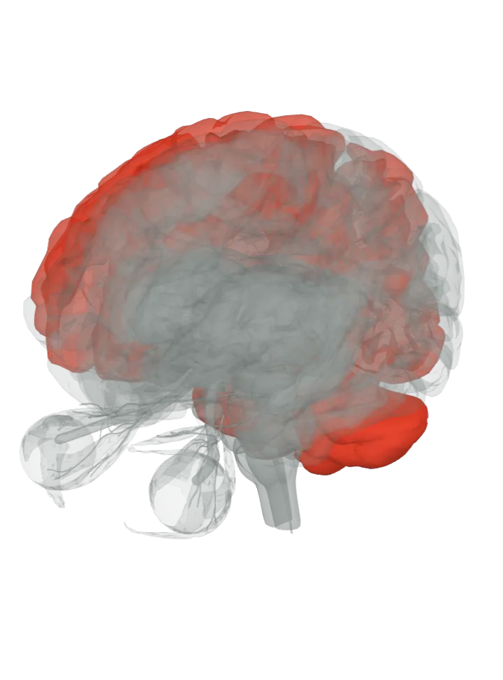
No single part: your eyes reach the target first and the hand follows. The frontal eye fields and the superior colliculus move your gaze, the visual cortex in the occipital lobe sends what you see up the ‘how’ stream to the superior parietal lobule, the premotor cortex prepares the reach, and the cerebellum keeps eye and hand in step. [1][2][3]
All 6 parts are lit together. Drag to turn, pinch or scroll to zoom, tap a part on the brain or a name in the list to focus it; .
The frontal eye fields, the premotor cortex and the visual cortex are regions on the lit parts, not separate shapes. The 3D model draws only whole gyri and lobes, so the part that holds each area is lit; the area’s exact border is not drawn.
How they work together
Gaze leads the hand: people who reached for a bar, grasped it and pressed a target switch with it looked at each grasp site and target before the fingertips or the bar got there, and never looked at their own hand or the moving bar. [1][11]
The eyes mark the spots the fingers go to: the moment gaze left a landmark matched the moment the hand finished its job there, as if each step was being checked off. [1]
Vision for action has its own route: the dorsal stream runs from the primary visual cortex to the posterior parietal cortex and guides reaching, apart from the ventral stream that recognises objects, in Goodale and Milner’s model. [2]
Where you look changes the network: reaching to a target in central vision used the medial intraparietal sulcus and the back of the dorsal premotor cortex, while reaching to one off to the side also drew on the parieto-occipital junction and a more forward part of the premotor cortex. [5]
Honest limit: these are networks seen in imaging and monkey recordings, not single centres; human scans show eye and hand movements lighting widespread, overlapping parietal and frontal areas, the superior colliculus’s reach cells are known from monkeys, and the frontal eye fields and premotor cortex are regions on lit gyri, so whole parts are lit, not exact borders. These sources tie the parts to looking and reaching, not to finger dexterity as a whole. [12][9][13]
Getting the eyes onto the target is the first half of the job: the eye movements page lights the frontal eye fields, the superior colliculus and the muscles and nerves that turn the eye. [6][8]What part of the brain controls eye movements?
Carrying out the reach is the motor system’s job: the movement page lights the premotor cortex with the primary motor cortex and the basal ganglia. [7]What part of the brain controls movement?
Facts describe anatomy and normal function only. A part lit here takes part in hand–eye coordination; it does not do it alone, and it does other jobs too. The 3D model is the adult male BodyParts3D brain (credits and licence); on it the brainstem includes the midbrain, pons and medulla, so lighting the brainstem lights all three.