brainlocation

For learning, not medical advice.

All parts of the brain › Functions › Disgust

What part of the brain controls disgust?

No single part, but the anterior insula comes closest: it is active both when you feel disgust, at a foul smell, and when you see disgust on someone else’s face, with the anterior cingulate cortex joining in more weakly. [1][2][3]

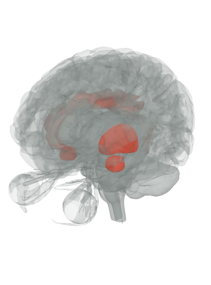
The insula, the putamen, the amygdala and the cingulate gyrus lit in red on a 3D model of the brain, seen from the front left and above

Loading the 3D brain…

Lit: whole parts; region borders not drawn.

Slice

All 4 parts are lit together. Drag to turn, pinch or scroll to zoom, tap a part on the brain or a name in the list to focus it; .

The putamen, in the basal ganglia, works with the insula in recognising disgust, and the amygdala takes part in some studies. The anterior insula is also a taste and smell area: disgust began as a reaction to offensive food. [1][2][3]

The anterior cingulate cortex is a region on a lit gyrus, not a separate shape. The 3D model draws only whole gyri, so the gyrus that holds each area is lit; the area’s exact border is not drawn.

How they work together

  • Disgust is a reaction of rejection or revulsion to something that might be contagious, or that is offensive, distasteful or unpleasant; it is thought to have evolved as a response to offensive foods that could cause harm. [3]
  • Feeling it and seeing it share circuitry: the same anterior insula sites were active when people smelled a disgusting odour and when they watched video of a disgusted face, which the authors read as the observer’s brain re-creating the emotion. [1]
  • The anterior insula is a smell and taste centre that also handles gut sensations and automatic body responses; electrically stimulating it during brain surgery brought on nausea and an uneasy stomach. [3]
  • Different disgusts, different patterns: contamination and mutilation pictures both drove the anterior insula, but only mutilation also drove the right superior parietal cortex, and strong disgust faces drew in a circuit through the striatum and thalamus as well. [4][6]
  • Honest limit: these are networks seen in imaging and lesion studies, not single centres; there is no one ‘disgust centre’, the amygdala shows up in some studies and not in others, and the anterior cingulate cortex is a region on a lit gyrus, so whole parts are lit, not exact borders. [6][5][7]

Disgust is one of the basic emotions with its own consistent pattern in imaging: the emotions page lights the amygdala, hypothalamus, cingulate gyrus, insula and prefrontal cortex. [8] What part of the brain controls emotions?

Disgust is literally ‘bad taste’, and the anterior insula also responds to offensive tastes: the taste page lights the insula with the taste pathway that reaches it. [6] What part of the brain controls taste?

Sources

  1. Wicker, Keysers, Plailly, Royet, Gallese & Rizzolatti 2003, Both of us disgusted in My insula: the common neural basis of seeing and feeling disgust (Neuron, PubMed abstract)
  2. Calder, Keane, Manes, Antoun & Young 2000, Impaired recognition and experience of disgust following brain injury (Nat Neurosci, PubMed abstract)
  3. Wikipedia: Disgust
  4. Wright, He, Shapira, Goodman & Liu 2004, Disgust and the insula: fMRI responses to pictures of mutilation and contamination (NeuroReport, PubMed abstract)
  5. Karama, Armony & Beauregard 2011, Film excerpts shown to specifically elicit various affects lead to overlapping activation foci in a large set of symmetrical brain regions in males (PLoS One, free in PMC)
  6. Phillips et al. 1997, A specific neural substrate for perceiving facial expressions of disgust (Nature, PubMed abstract)
  7. BodyParts3D (DBCLS), the atlas this 3D model is built from
  8. Vytal & Hamann 2010, Neuroimaging support for discrete neural correlates of basic emotions: a voxel-based meta-analysis (J Cogn Neurosci, PubMed abstract)

Facts describe anatomy and normal function only. A part lit here takes part in disgust; it does not do it alone, and it does other jobs too. The 3D model is the adult male BodyParts3D brain (credits and licence); on it the brainstem includes the midbrain, pons and medulla, so lighting the brainstem lights all three.