The vestibular nuclei are the brainstem nuclei of the vestibular nerve, the balance half of the vestibulocochlear nerve: four nuclei on the floor of the fourth ventricle, grouped in both the pons and the medulla oblongata. They take the inner ear’s signals about how the head tilts and moves and pass them on to the eye-muscle nuclei, the spinal cord and the cerebellum. [1][2]
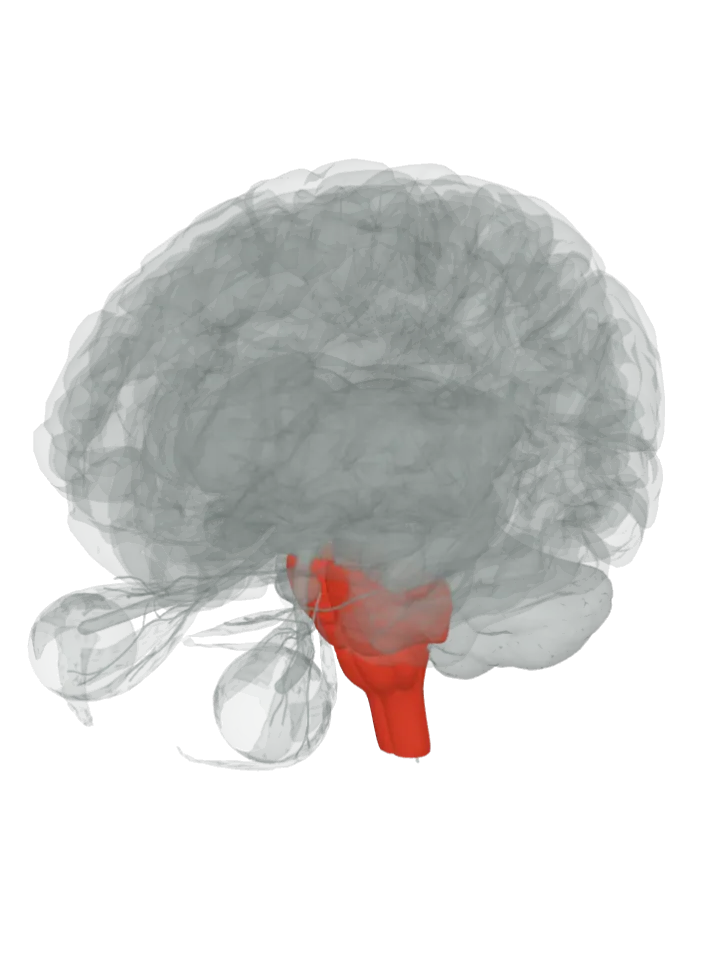
The pons and medulla oblongata are lit together; the vestibular nuclei are not drawn on their own. The side (sagittal) slice button cuts through them. Drag to turn, pinch or scroll to zoom, tap any part to name it; .
Not a separate shape in this atlas; lit: the part that holds it. Lit: the whole pons and medulla oblongata. Terminologia Anatomica groups the vestibular nuclei in both the pons and the medulla oblongata, under the floor of the fourth ventricle; the medial nucleus, for one, is in the medulla. So both parts are lit together. They have no shape of their own in the BodyParts3D model, so nothing is drawn at a guessed point. [1][3]
Lit here: the pons and the medulla oblongata, the parts that hold the vestibular nuclei — see each on its own page.
There are four: the superior (Bechterew’s), lateral (Deiters’), medial (Schwalbe’s) and inferior vestibular nuclei, all at the floor of the fourth ventricle. [1][4][5][3][6]
The superior nucleus is the upper outer (dorso-lateral) part of the group; the lateral nucleus continues upward and outward from the principal nucleus; the medial nucleus is in the medulla oblongata. [4][5][3]
The vestibular nerve’s fibres enter the medulla on the inner side of the cochlear fibres, pass between the inferior cerebellar peduncle and the spinal tract of the trigeminal nerve, then split into ascending and descending branches that end in the nuclei. [1]
Through the vestibulospinal tract they adjust muscle tone and the position of the limbs and head to hold posture and keep the body balanced: the lateral tract, from Deiters’ nucleus, runs down the same side to the lower (lumbar and sacral) spinal cord; the medial tract runs on both sides to the neck segments for head and neck movements. [2][3]
Fibres from the medial and lateral nuclei climb the medial longitudinal fasciculus to the nuclei of the oculomotor nerve, the route of the vestibulo-ocular reflex that keeps the eyes on target while the head moves; the superior nucleus sends uncrossed fibres the same way to the oculomotor and trochlear nuclei. [1][4][7]
Connections
Some axons of the lateral nucleus, and possibly the medial, run up through the inferior cerebellar peduncle to the roof nuclei of the cerebellum on the opposite side; other vestibular nerve fibres go straight to the cerebellum without stopping in the nuclei. [1]
Other names
Also the vestibular nucleus or VN; three carry the names of anatomists: Bechterew’s nucleus (superior), Deiters’s nucleus (lateral) and the Schwalbe nucleus (medial). [1][4][5][3]
Size
The model has no shape for the vestibular nuclei itself, so it is not measured here. The pons that holds it measures about 34 mm front to back, 29 mm top to bottom and 42 mm side to side on this model (the box it fits in, not its volume). [8]
The model has no shape for the vestibular nuclei itself, so it is not measured here. The medulla oblongata that holds it measures about 33 mm front to back, 41 mm top to bottom and 27 mm side to side on this model (the box it fits in, not its volume). [8]
Facts describe anatomy and normal function only. The 3D model is the adult male BodyParts3D brain (credits and licence); real brains vary in size and shape, so its measurements describe one model, not anyone’s brain.