How does the vestibulo-ocular reflex keep the eyes steady?
It keeps your eyes on target while your head moves: the inner ear senses the turn, the vestibular nuclei in the brainstem pass it on, and eye muscles turn both eyes the opposite way, lagging the head by under 10 ms. [1]
The atlas has no shape for the vestibular nerve, its nuclei or the medial longitudinal fasciculus; lit: the parts it runs through, in order.
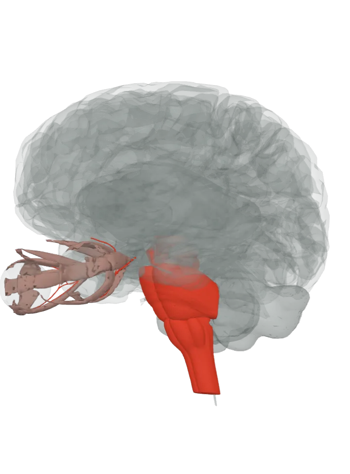
The brain starts seen from the left, a little from the front and below, so the brainstem and the eyes in front of it both show. Step through lights one step at a time, about 1.2 s each, from the inner ear’s nerve to the eye muscles; tap a step below to light it. The lit step is in the bright highlight colour, the steps before it stay lit in a paler tint of it and the steps still ahead are grey. Drag to turn, pinch or scroll to zoom. Step through is off because your device asks for reduced motion; tap each step instead.
The vestibular nerve, the vestibular and abducens nuclei, the abducens nerve and the medial longitudinal fasciculus are not drawn, so their steps light the pons and medulla that hold them. The oculomotor nerve is drawn, and the eye muscles light in their own colour with the last step. [2][1]
The route, in 6 steps
When the head turns, the semicircular canals of the inner ear sense the rotation. The signal runs through the vestibular ganglion and along the vestibular part of the vestibulocochlear nerve into the brainstem, where the pons meets the medulla. The nerve has no shape in this atlas; lit: the pons and medulla oblongata at whose border it enters. [1][3]Where is the vestibular nerve (CN VIII)?
The nerve ends in the vestibular nuclei, on the floor of the fourth ventricle where the pons meets the medulla. From these nuclei, fibres cross the midline to the abducens nucleus of the other side. Not a separate shape in this atlas; lit: the parts that hold them, the pons and the medulla oblongata. [1][4]Where is the pons?Where is the medulla oblongata?
The abducens nucleus lies under the fourth ventricle in the lower pons, near the midline. Its motor neurons send the abducens nerve, which leaves where the pons meets the medulla, to the lateral rectus, the muscle that turns that eye outward. Neither has a shape in this atlas; lit: the pons and medulla oblongata, as on the abducens nerve’s own page. [5][1][6]Where is the abducens nucleus and nerve (CN VI)?
Lights nothing new: the medial longitudinal fasciculus has no shape in this atlas, so no line is drawn for it and the pons it climbs through stays lit. Interneurons of the abducens nucleus send their axons across the midline and up this bundle to the oculomotor nucleus of the other side, so the second eye turns with the first; it also carries fibres from the vestibular nuclei to the oculomotor and trochlear nuclei. [7][5][1]
The oculomotor nucleus lies in the grey matter of the midbrain, under the cerebral aqueduct. Its motor neurons send the oculomotor nerve forward to the medial rectus, which turns the eye inward toward the nose. The nucleus is not a separate shape in this atlas; lit: the part that holds it, the midbrain, and the oculomotor nerve, which the atlas does draw. [8][1][6]Where is the midbrain?Where is the oculomotor nerve?
The lateral rectus of one eye and the medial rectus of the other contract together, turning both eyes as far as the head turned but the opposite way, so the image holds still on each retina. The abducens nerve that works the lateral rectus has no shape here, so the oculomotor nerve stays lit beside the muscles. [1][6]Where are the extraocular muscles?
Lit with it: the six muscles of each eye, in their own colour, as the atlas draws them; for a turn to the side, the lateral rectus of one eye and the medial rectus of the other do the work. [2][6][1]
More about the vestibulo-ocular reflex
Other names: VOR, vestibulo-ocular reflex, vestibuloocular reflex [1]
It is one of the fastest reflexes in the body: the signal from the semicircular canals reaches the eye muscles over a chain of only three neurons, and the eyes lag the head by less than 10 ms. [1]
It does not depend on what is seen: it runs on head movement sensed by the inner ear, so it works in total darkness and with the eyes closed. [1]
Ideally the eyes turn exactly as far as the head, a gain of 1.0. The steps above trace the side-to-side turn; similar pathways serve the up-and-down and rolling (torsional) parts of the reflex. [1]
The same vestibular nuclei also drive the vestibulospinal tract, which keeps the body upright and the head steady; both serve balance. [4][9]
Facts describe anatomy and normal function only. The 3D model is the adult male BodyParts3D brain (credits and licence); it has no shape for tracts, so a pathway is shown by the parts it runs through.