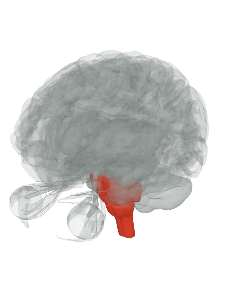
The cochlear nuclei are the two brainstem nuclei where every hearing fibre from the cochlea ends: the ventral and the dorsal cochlear nucleus, on the back and outer (dorsolateral) side of the brainstem, spanning the junction of the pons and the medulla oblongata. All sound information enters the brain through them, and its processing begins there. [1]
The pons and medulla oblongata are lit together; the cochlear nuclei are not drawn on their own. The side (sagittal) slice button cuts through them. Drag to turn, pinch or scroll to zoom, tap any part to name it; .
Not a separate shape in this atlas; lit: the part that holds it. Lit: the whole pons and medulla oblongata. Wikipedia’s Cochlear nucleus article places them at the dorsolateral side of the brainstem, spanning the junction of the pons and medulla, so both parts are lit together. The ventral nucleus lies ventrolateral to the inferior cerebellar peduncle; the dorsal nucleus curves over it and wraps around the peduncle. They have no shape of their own in the BodyParts3D model, so nothing is drawn at a guessed point. [1]
Lit here: the pons and the medulla oblongata, the parts that hold the cochlear nuclei — see each on its own page.
There are two: the ventral cochlear nucleus (VCN), unlayered, which the nerve root divides into an anteroventral and a posteroventral part, and the dorsal cochlear nucleus (DCN, the acoustic tubercle), layered and cortex-like, on the dorso-lateral surface of the brainstem. [1][2]
All auditory nerve fibres from the cochlea make their first synapses here, carrying sound from the ear on the same side of the head. [1][2]
They keep the cochlea’s map of pitch (tonotopic organisation): fibres tuned to low frequencies end in the ventral and lateral parts, fibres tuned to high frequencies in the dorsal parts, and middle frequencies in between. [1][3]
The dorsal nucleus takes input from the ventral nucleus as well as from the nerve, so it is in a sense a second-order relay; in the ventral nucleus, bushy cells receive the nerve through unusually large endings, the end bulbs of Held. [1][3]
Connections
Input: the cochlear nerve, the hearing half of the vestibulocochlear nerve, enters at the nerve root in the ventral nucleus, and its fibres branch to both nuclei. [1][3]
Output: three fibre bundles, mostly to the opposite side: the ventral acoustic stria (the trapezoid body) to the superior olive of both sides, and the dorsal and intermediate acoustic striae, which climb to the lateral lemniscus. [1]
Other names
Also the cochlear nucleus or cochlear nuclear complex (CN); the dorsal nucleus is also called the tuberculum acusticum (acoustic tubercle). [1][2]
Size
The model has no shape for the cochlear nuclei itself, so it is not measured here. The pons that holds it measures about 34 mm front to back, 29 mm top to bottom and 42 mm side to side on this model (the box it fits in, not its volume). [4]
The model has no shape for the cochlear nuclei itself, so it is not measured here. The medulla oblongata that holds it measures about 33 mm front to back, 41 mm top to bottom and 27 mm side to side on this model (the box it fits in, not its volume). [4]
Facts describe anatomy and normal function only. The 3D model is the adult male BodyParts3D brain (credits and licence); real brains vary in size and shape, so its measurements describe one model, not anyone’s brain.