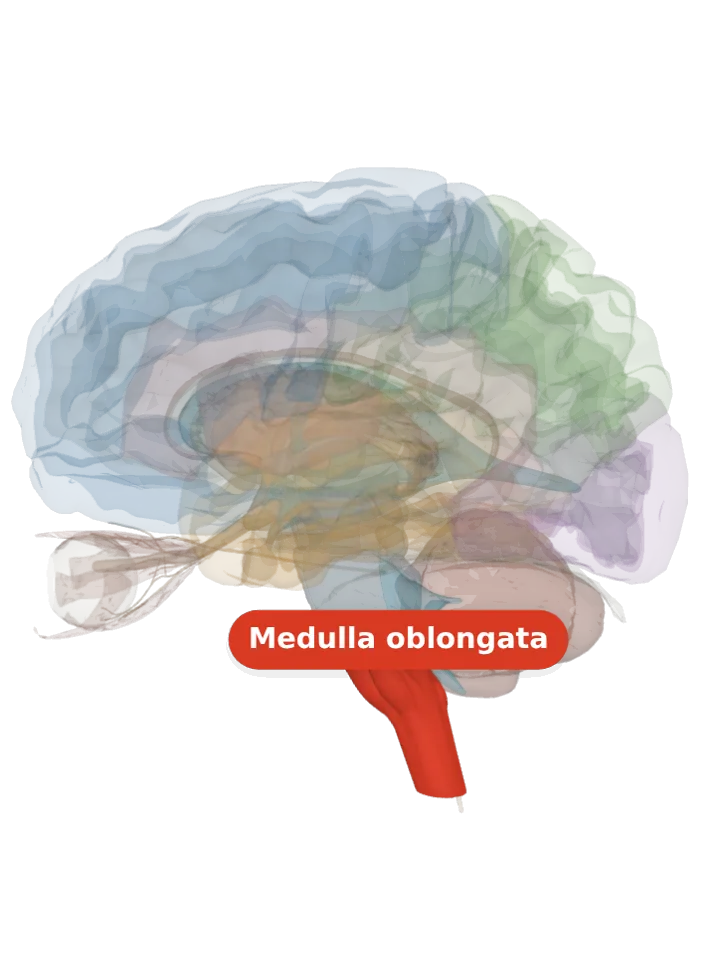
The dorsal vagal nucleus (the dorsal nucleus of the vagus nerve) is a cranial nerve nucleus in the medulla oblongata, just beneath the floor of the fourth ventricle. It holds the parasympathetic nerve cells whose fibres run out in the vagus nerve to the gut, the lungs and the other organs of the chest and abdomen. [1]
Opens cut by a side (sagittal) slice through the medulla oblongata, which holds the dorsal vagal nucleus. Drag to turn, pinch or scroll to zoom, tap any part to name it; .
Not a separate shape in this atlas; lit: the part that holds it. Lit: the whole medulla oblongata. The dorsal vagal nucleus lies in the medulla oblongata, ventral to (just in front of) the floor of the fourth ventricle. It is not in the pons, so only the medulla is lit. It has no shape of its own in the BodyParts3D model, so nothing is drawn at a guessed point. [1]
Lit here: the medulla oblongata, the part that holds the dorsal vagal nucleus — see it on its own page.
It sits in the medulla oblongata, ventral to (just in front of) the floor of the fourth ventricle. [1]
It lies just beside the hypoglossal nucleus, the motor nucleus of the tongue, which raises the hypoglossal trigone in the floor of the fourth ventricle. [2][1]
Its parasympathetic nerve cells give the vagus nerve its parasympathetic supply to the gastrointestinal tract and the lungs, as well as to other organs of the chest and abdomen. [1]
Among the jobs of that outflow are narrowing the airways (bronchoconstriction) and making glands secrete. [1]
The vagus nerve’s fibres to the heart mostly start elsewhere: their parasympathetic cell bodies sit in the nucleus ambiguus; together the two nuclei slow the heart when blood pressure rises quickly. [1][4]
Connections
Input from the solitary nucleus, which passes on what the vagus and glossopharyngeal nerves sense from the organs. [5]
Output: preganglionic parasympathetic fibres that leave the medulla in the vagus nerve and end on nerve cells (ganglia) in or near the organs they serve. [1][3]
Other names
Also the dorsal nucleus of the vagus nerve, the posterior nucleus of the vagus nerve, the dorsal motor nucleus of the vagus, or in Latin nucleus dorsalis nervi vagi or nucleus posterior nervi vagi; “dorsal” because it lies towards the back of the medulla. [1][4]
Size
The model has no shape for the dorsal vagal nucleus itself, so it is not measured here. The medulla oblongata that holds it measures about 33 mm front to back, 41 mm top to bottom and 27 mm side to side on this model (the box it fits in, not its volume). [6]
Facts describe anatomy and normal function only. The 3D model is the adult male BodyParts3D brain (credits and licence); real brains vary in size and shape, so its measurements describe one model, not anyone’s brain.