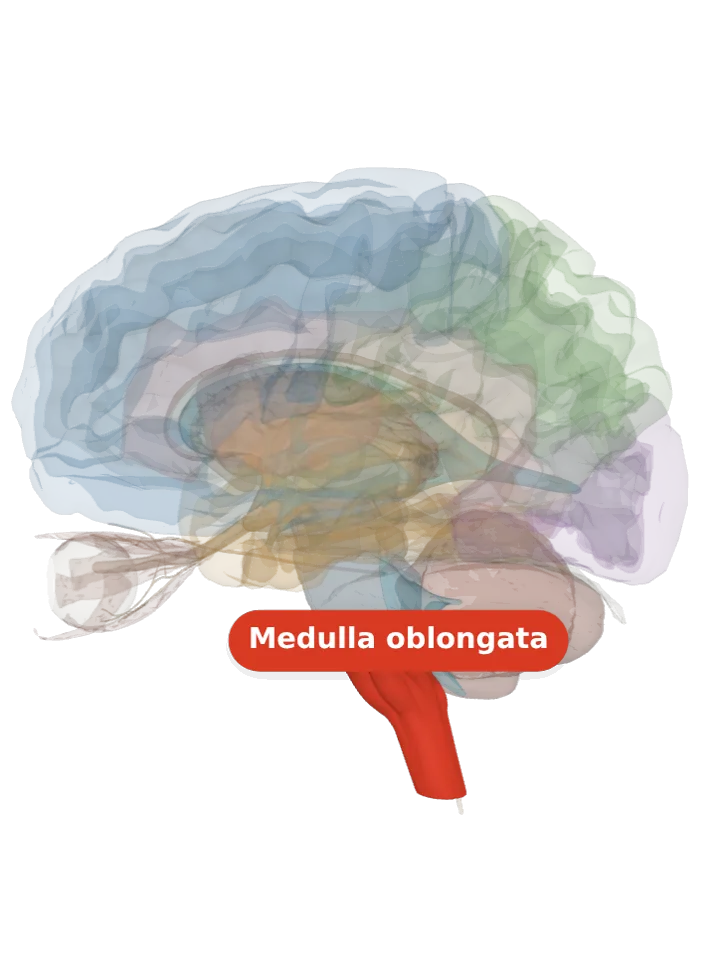
The solitary nucleus (the nucleus of the solitary tract) is a roughly vertical column of grey matter in the medulla oblongata, wrapped around a bundle of incoming fibres called the solitary tract. It is the brainstem’s first stop for taste and for signals from the heart, lungs, airways and gut, which reach it through the facial, glossopharyngeal and vagus nerves. [1][2]
Opens cut by a side (sagittal) slice through the medulla oblongata, which holds the solitary nucleus. Drag to turn, pinch or scroll to zoom, tap any part to name it; .
Not a separate shape in this atlas; lit: the part that holds it. Lit: the whole medulla oblongata. The solitary nucleus runs lengthwise through the medulla oblongata, in its back and outer (posterolateral) part, around the solitary tract, which carries on down to the top of the spinal cord. The column is in the medulla, so the pons above it is not lit. It has no shape of its own in the BodyParts3D model, so nothing is drawn at a guessed point. [1][2]
Lit here: the medulla oblongata, the part that holds the solitary nucleus — see it on its own page.
It is a roughly vertical column of grey matter in the medulla oblongata. Its nerve cells’ axons make up most of the solitary tract, the compact fibre bundle it surrounds, which runs lengthwise through the back and outer (posterolateral) part of the medulla. [1][2]
It is laid out by what it serves along its length: the taste cells, called the gustatory nucleus, sit in the upper (rostral) part, and the cells that take signals from the heart, blood vessels, lungs and gut sit in the lower (caudal) part. Across its width it is divided into dorsomedial, dorsolateral and ventrolateral subnuclei. [1]
The tract it surrounds, also called the tractus solitarius, descends to the upper segments of the spinal cord in the neck. [2]
What it does
It receives taste: from the front two-thirds of the tongue through the facial nerve (its chorda tympani branch), from the back third through the glossopharyngeal nerve and from the epiglottis through the vagus nerve. [1]
It also receives the body’s inner signals: blood pressure and blood chemistry from the carotid sinus and carotid body (through the glossopharyngeal nerve) and from the aortic arch (through the vagus), and stretch and chemical signals from the heart, lungs, airways, gut and liver. [1]
Its first neurons can run simple automatic reflexes themselves: the baroreflex that steadies blood pressure, the cough reflex, breathing reflexes and the reflexes that set the movement and secretions of the gut all pass through it. [1]
In the gag reflex it is the middle step: the glossopharyngeal nerve’s signal from the throat ends here, and the solitary nucleus passes it to the nucleus ambiguus, whose motor fibres answer through the glossopharyngeal and vagus nerves. [1][3]
It also projects to other cranial nerve nuclei: the salivatory nuclei, the hypoglossal nucleus, the dorsal nucleus of the vagus nerve and the nucleus ambiguus. [1]
Other names
Also the nucleus of the solitary tract, nucleus solitarius or nucleus tractus solitarii (Latin), shortened to SN; its taste part is the gustatory nucleus. “Solitary” comes from the solitary tract it surrounds. [1]
Size
The model has no shape for the solitary nucleus itself, so it is not measured here. The medulla oblongata that holds it measures about 33 mm front to back, 41 mm top to bottom and 27 mm side to side on this model (the box it fits in, not its volume). [4]
Facts describe anatomy and normal function only. The 3D model is the adult male BodyParts3D brain (credits and licence); real brains vary in size and shape, so its measurements describe one model, not anyone’s brain.