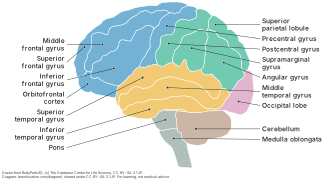
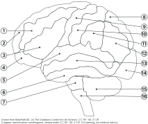
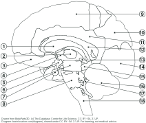
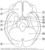
Three standard views drawn from the 3D brain model this site uses: each coloured shape is the outline of one real part of the model, and each label points into its own shape. Tap a name to see that part in 3D. Printing puts one view on each A4 or Letter page.
Drawn from BodyParts3D, (c) The Database Center for Life Science, CC BY-SA 2.1 JP. Diagram: brainlocation.com/diagram/, shared under CC BY-SA 2.1 JP (credits). For learning, not medical advice.
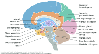
Midline view: the brain cut down the middle
Medial (midsagittal) view, inner face of the right hemisphere; front of the brain to the left. 18 parts labelled.
Drawn from BodyParts3D, (c) The Database Center for Life Science, CC BY-SA 2.1 JP. Diagram: brainlocation.com/diagram/, shared under CC BY-SA 2.1 JP (credits). For learning, not medical advice.
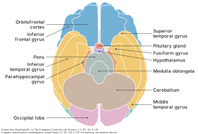
Underside view: the base of the brain
Inferior view, both hemispheres; front of the brain at the top, the left hemisphere on the right of the sheet. 13 parts labelled.
Drawn from BodyParts3D, (c) The Database Center for Life Science, CC BY-SA 2.1 JP. Diagram: brainlocation.com/diagram/, shared under CC BY-SA 2.1 JP (credits). For learning, not medical advice.
How it is drawn
Every shape is computed from the same BodyParts3D meshes the 3D brain on this site shows, not traced by hand. Each view projects the meshes straight onto the page (an orthographic projection, so nothing is shrunk by perspective) on a grid of 0.25 mm cells, and each cell keeps whichever part is nearest the eye.
For the side and midline views the model is first cut in two at the midline. The side view keeps the left half and looks at it from the left; the midline view keeps the right half and looks at its cut face from the same side, so in both the front of the brain is on the left of the sheet. The underside view looks up at the whole brain from below, front at the top; as on any view from below, the brain’s left half appears on the right of the sheet.
Each part’s cells are traced into an outline and smoothed to within about 0.35 mm. Every leader line starts at the point deepest inside its own shape, so it never points at a border or at a neighbour; test/diagram.test.cjs re-projects the model and checks this for every label on every sheet.
Sizes are true to the model: in the side view the brain is about 180 mm from front to back. Measure it on a print and the ratio to the sheet tells you the scale.
What it does not show
Only what the model has a shape for. The atlas has no septum pellucidum, the thin wall between the two lateral ventricles, so on the midline view you look straight into the right lateral ventricle under the corpus callosum. It has no fornix, mammillary bodies, olfactory bulbs or cranial nerves either, so none of them is drawn or labelled.
On the inner face, the model’s superior parietal lobule shape runs on over the area usually called the precuneus, and its occipital lobe shape covers the cuneus and the lingual gyrus. The model has no separate shapes for those three, so they are labelled with the shape’s own name.
Thin parts seen edge-on (the fourth ventricle beside the cerebellum, the hypothalamus under the thalamus) come out as small or ragged patches; where a piece of a part was smaller than about 7.5 mm² on the page it was folded into the part around it.
It is one adult brain. Real brains differ in size and in exactly where one gyrus ends, so treat the borders as a typical layout, not a measurement of anyone.
Printing and sharing
Print from the browser (or the button above): each view gets its own page and fits A4 and US Letter in portrait, with the colour key and the credit line on the sheet. Choose “black and white” for the blank worksheet; the labelled sheets read best in colour.
The drawings are derived from BodyParts3D, so they carry its licence, CC BY-SA 2.1 Japan: you may copy, hand out, edit and reuse them, also commercially, as long as the credit line stays on and anything you make from them is shared under the same licence. Details on the credits page.