The occipitotemporal sulcus, also called the lateral occipitotemporal sulcus, is a long groove on the underside of each hemisphere. It runs from near the occipital pole at the back to a short distance from the temporal pole in front, and separates the inferior temporal gyrus on its outer side from the fusiform gyrus on its inner side. [1][2]
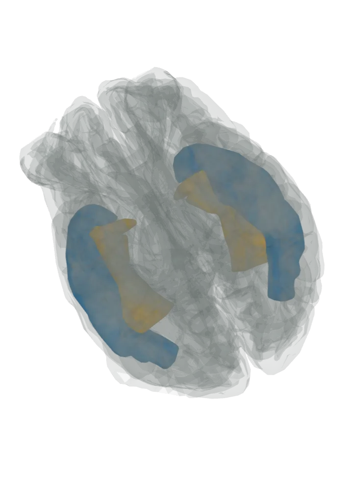
Seen from below: on the underside of each hemisphere the inferior temporal gyrus is lit in blue along the outer edge, the fusiform gyrus in orange inside it; the brainstem and cerebellum sit between the two halves. The groove runs where blue meets orange; the top (axial) slice button cuts across it. Drag to turn, pinch or scroll to zoom, tap any part to name it; .
A groove has no shape of its own; lit: the parts on either side of it. Lit: on the outer side, the inferior temporal gyrus in blue; on the inner side, the fusiform gyrus in orange. The occipitotemporal sulcus separates the fusiform gyrus from the inferior temporal gyrus on its outer (lateral) side; the collateral sulcus bounds the fusiform gyrus on its inner side. It runs on the underside of the brain, so this page opens seen from below. [2][1]
Where it is
It crosses the underside of the temporal lobe, which is concave and continues at the back into the underside of the occipital lobe; it runs from near the occipital pole to within a short distance of the temporal pole, and is often interrupted by bridging gyri. [1]
The inferior temporal gyrus runs along the bottom of the outer side of the temporal lobe and wraps round its lower edge onto the underside, where this groove limits it. [3]
A shallower groove, the mid-fusiform sulcus, splits the fusiform gyrus along its length; the lateral part of the fusiform gyrus lies between the mid-fusiform sulcus and this groove. [2]
What it does
A fold, not a working part. Both banks are high in the ventral stream of vision: the inferior temporal gyrus is associated with representing objects, places, faces and colours, and the fusiform gyrus has been linked with recognition. [3][2]
The visual word form area, a functional region thought to identify letters and words from their shapes during reading, is in the left fusiform gyrus and the cortex around it. [4]
Other names
It is also called the lateral occipitotemporal sulcus; the collateral sulcus, on the other side of the fusiform gyrus, is the medial occipitotemporal sulcus, and the fusiform gyrus between them is also called the lateral occipitotemporal gyrus. [1][5][2]
Not to be confused with the inferior temporal sulcus, the groove on the outer side of the temporal lobe that separates the middle temporal gyrus from the inferior temporal gyrus below it. [3]
Size
The model has no shape for the occipitotemporal sulcus, so the groove itself is not measured here. Measured from the model instead: on the left hemisphere, the stretch where the inferior temporal gyrus and the fusiform gyrus come within 3 mm of each other fits in a box about 65 mm front to back, 24 mm top to bottom and 28 mm side to side (the box that stretch fits in, not the groove’s length along its bends or its depth). [6]
Facts describe anatomy and normal function only. The 3D model is the adult male BodyParts3D brain (credits and licence); real brains vary in size and shape, so its measurements describe one model, not anyone’s brain.