The inferior frontal sulcus is the groove that runs front to back on the outer side of each frontal lobe, between the middle frontal gyrus above and the inferior frontal gyrus below. Its back end meets the inferior precentral sulcus, at a spot called the inferior frontal junction. [1][2]
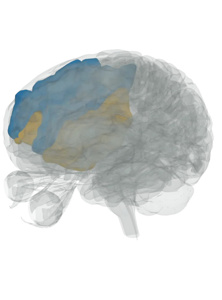
Seen from the left and in front: the middle frontal gyrus is lit in blue, the inferior frontal gyrus in orange, on both hemispheres. The groove runs where blue meets orange; the front (coronal) slice button cuts across it. Drag to turn, pinch or scroll to zoom, tap any part to name it; .
A groove has no shape of its own; lit: the parts on either side of it. Lit: above, the middle frontal gyrus in blue; below, the inferior frontal gyrus in orange. The inferior frontal sulcus lies between the middle frontal gyrus and the inferior frontal gyrus; it is the upper border of the inferior frontal gyrus and the lower border of the middle frontal gyrus. [1][3][4]
Its back end meets the inferior precentral sulcus; the area at that meeting point is called the inferior frontal junction. [2][5]
What it does
A fold, not a working part. The inferior frontal junction area, where it meets the inferior precentral sulcus, is involved in working memory and attention; in a meta-analysis of imaging studies, oddball and attention, working-memory, task-switching and Stroop tasks all activated that junction. [2][5]
Below it, the inferior frontal gyrus holds the pars opercularis and pars triangularis, which together make up Broca’s area. [3]
The model has no shape for the inferior frontal sulcus, so the groove itself is not measured here. Measured from the model instead: on the left hemisphere, the stretch where the middle frontal gyrus and the inferior frontal gyrus come within 3 mm of each other fits in a box about 65 mm front to back, 50 mm top to bottom and 23 mm side to side (the box that stretch fits in, not the groove’s length along its bends or its depth). [7]
Facts describe anatomy and normal function only. The 3D model is the adult male BodyParts3D brain (credits and licence); real brains vary in size and shape, so its measurements describe one model, not anyone’s brain.