How they work together
- There are three kinds of tears. Basal tears keep the eye wet at about 0.5–2.2 µL a minute; irritation can raise the rate up to about 100-fold, to some 300 µL a minute (reflex tears); emotional (psychic) tears are the ones shed in emotional states. In a day a person makes about 0.75 to 1.1 grams of tears, less with age. [2][5]
- Reflex and emotional tears enter the circuit by different roads: when the trigeminal nerve is cut, reflex tears stop but emotional tears do not. Both end on the same parasympathetic route, which works the tear gland with acetylcholine. [2]
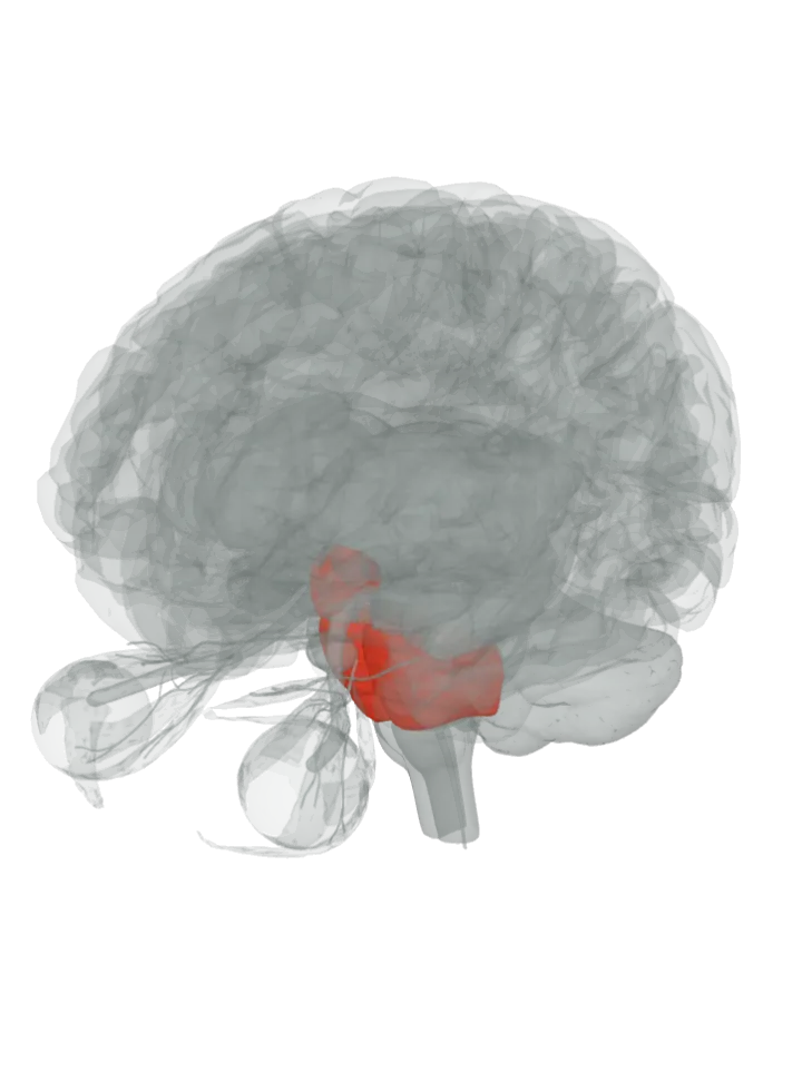
- Anatomical studies in animals find inputs to the superior salivatory nucleus from the frontal cortex, basal ganglia, thalamus and paraventricular hypothalamus, as well as from the trigeminal nerve. Only the hypothalamus is lit, because it is the one input whose link to tearing has been tested directly; the others are known only from anatomy. [3]
- Emotional tears carry larger amounts of some hormones, including prolactin, adrenocorticotropic hormone and leu-enkephalin, and of potassium and manganese. [5]
- Honest limit: the oxytocin route was shown in mice, in a 2022 preprint, and emotional tearing is often assumed to be unique to humans, so it is a lead, not the full human circuit. The page covers how tears are made and sent, not the crying face, voice or breathing, and nothing about any one person. [3]
Laughter, the other loud emotional outburst, has its own page; it lights the hypothalamus and pons as well, with the brainstem’s periaqueductal grey and the cingulate gyrus. [6] What part of the brain controls laughter?
The same facial nerve works the face muscles of a crying face: the facial expressions page lights the motor cortex and the pons, where the facial motor nucleus sits. [1] What part of the brain controls facial expressions?