brainlocation

For learning, not medical advice.

All parts of the brain › Functions › Social behaviour

What part of the brain controls social behaviour?

No single part: reading other people draws on a set of regions. The medial and ventromedial prefrontal cortex are active when you think about what others feel and intend, the temporoparietal junction when you work out what someone else believes, the superior temporal sulcus reads movement, gaze and voices, the amygdala gives a social signal its emotional weight and the fusiform gyrus picks out faces. [1][2][3]

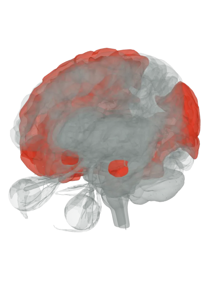
The superior frontal gyrus, the orbitofrontal cortex, the supramarginal gyrus, the angular gyrus, the superior temporal gyrus, the amygdala and the fusiform gyrus lit in red on a 3D model of the brain, seen from the front left and above

Loading the 3D brain…

Lit: whole parts; region borders not drawn.

Slice

All 7 parts are lit together. Drag to turn, pinch or scroll to zoom, tap a part on the brain or a name in the list to focus it; .

The ventromedial prefrontal cortex is a region on a lit part, not a separate shape. The 3D model draws only whole gyri and lobes, so the part that holds each area is lit; the area’s exact border is not drawn.

The temporoparietal junction is a region on the angular gyrus, not a separate shape. The 3D model has no shape for it, so the whole angular gyrus is lit; the region’s exact border is not drawn.

The superior temporal sulcus is a region on the superior temporal gyrus, not a separate shape. The 3D model has no shape for it, so the whole superior temporal gyrus is lit; the region’s exact border is not drawn.

How they work together

  • Roughly, the parts fall into two groups. The fusiform gyrus, superior temporal sulcus and amygdala take in social signals: faces, bodies moving and voices. The medial prefrontal cortex and the TPJ are active when you think about what another person believes or intends, with the posterior cingulate cortex, another region active in theory-of-mind tasks, working in step with the medial prefrontal cortex even at rest. [1]
  • The first clue came from Phineas Gage, a railroad construction foreman whose behaviour was reported to change after an 1848 accident damaged one or both of his frontal lobes; later work found that frontal damage can change emotional responses to social stimuli and performance on theory-of-mind tasks. [4][6]
  • The human brain does much of its growing among other people: at birth it is about 25% of its adult volume, against nearly 50% for a chimpanzee and about 70% for a macaque, and the prefrontal cortex, tied to decision-making and social behaviour, expanded greatly in humans compared with other primates. [1][4]
  • Honest limit: these regions come mostly from brain imaging and from what changes after damage, and none of them is only social (the fusiform face area also responds to objects people are expert in; the amygdala tracks arousal in general). The atlas lights whole gyri, not the exact patches, and the page describes how social perception works in general, nothing about any one person. [1][3]

Sharing what someone else feels is its own page: empathy lights the insula, which is active when you watch someone you love receive a shock, with the cingulate gyrus and the same amygdala and TPJ. [1][2] What part of the brain controls empathy?

Telling who a face belongs to has its own page, lighting the fusiform face area with the occipital lobe and inferior temporal gyrus that feed it. [5] What part of the brain recognises faces?

Sources

  1. Adolphs (2009), The social brain: neural basis of social knowledge, Annual Review of Psychology 60:693–716 (free in PMC)
  2. Wikipedia: Temporoparietal junction
  3. Wikipedia: Superior temporal sulcus
  4. Wikipedia: Social cognition
  5. Wikipedia: Fusiform face area
  6. Wikipedia: Phineas Gage

Facts describe anatomy and normal function only. A part lit here takes part in social behaviour; it does not do it alone, and it does other jobs too. The 3D model is the adult male BodyParts3D brain (credits and licence); on it the brainstem includes the midbrain, pons and medulla, so lighting the brainstem lights all three.