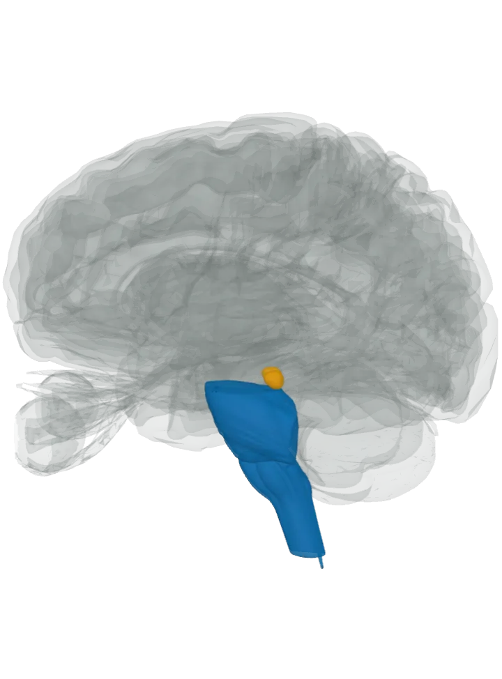
Made in: lit in blue
- Spinal cord, in the spinal cord (central canal): Its grey matter holds inhibitory interneurons that release glycine, among them the Renshaw cells, which feed back onto the motor neurons that drive the muscles; a mouse study found GlyT2, the pump that marks glycine neurons, in the spinal cord and the hindbrain. The atlas draws the cord only as its central canal, cropped just below the medulla, so that thin line is what is lit. [4][3][1]
- Medulla oblongata, lit as the medulla oblongata: In its pre-Bötzinger complex, on the ventral side of the lower medulla, the network that sets the breathing rhythm, about half of the interneurons are inhibitory, using glycine or GABA; the same mouse study found GlyT2 neurons in the medulla. The complex has no shape here, so the medulla is lit. [5][3]
- Superior olivary complex, lit as the pons: Its medial nucleus of the trapezoid body (MNTB) is made of round neurons that use glycine. The complex sits in the pons and has no shape of its own here, so the pons is lit; the mouse study also found GlyT2 neurons in the pons. [6][3]
Sent to: lit in orange
- Inferior colliculus (LSO → inferior colliculus): The midbrain’s hearing relay. The lateral superior olive’s fibres to the colliculus on their own side are mainly inhibitory, using glycine; those to the other side excite. [6]
- Not lit as sources: the midbrain and the cerebellum, where the same mouse study also found neurons carrying GlyT2; it places these neurons predominantly in the caudal regions of the brain and in the spinal cord. [3]
- Not lit as targets: the hippocampus, some of whose neurons carry glycine receptors beside GABA-A receptors, and the retina, where the receptor is especially common; the lit sources do not send glycine there. [2][1]
The pathways, as the sources name them
- Recurrent inhibition, from the Renshaw cells back onto the motor neurons that drive them, inside the lit spinal cord. A branch of a motor neuron’s own axon excites a Renshaw cell, which releases glycine back onto that motor neuron and others of its pool: a negative feedback loop. [4] See What part of the brain controls movement?
- MNTB → LSO, from the medial nucleus of the trapezoid body to the lateral superior olive, both inside the lit pons. The MNTB, driven by the cochlear nucleus of the other side, inhibits the LSO with glycine; the LSO weighs that against excitation from its own side’s ear, the basis of telling which ear hears a sound louder. [6] See What part of the brain controls hearing?
- LSO → inferior colliculus, from the lateral superior olive in the pons up to the inferior colliculus of the midbrain. The projection to the same side is primarily inhibitory and glycinergic, the one to the other side excitatory. [6] See What part of the brain controls hearing?
- Inhibition in the breathing network, between the neurons of the pre-Bötzinger complex, inside the lit medulla. Blocking the glycine and GABA inhibition there leaves its neurons unable to switch from one phase of the rhythm to the next as they should, seen in living animals as shorter breaths in. [5] See What part of the brain controls breathing?
Good to know
- Not only a brake: glycine is a required co-agonist, with glutamate, at the excitatory NMDA receptors. Glycine transporter 1, mainly on glial cells throughout the brain and spinal cord, clears it from those synapses; glycine transporter 2, on neurons of the hindbrain and spinal cord, from the inhibitory ones. [1]
- How the brake works: the glycine receptor is a channel of five subunits around a central pore. When glycine binds, chloride flows into the cell and pushes it further from firing; the adult receptor is the α1β form. [2]
- Not always a brake: in the embryo glycine excites, because chloride’s equilibrium potential is then more positive; the embryonic receptor is made of five α2 subunits. [2]
- The simplest amino acid: its side chain is a single hydrogen atom, and cells build it from serine with the enzyme serine hydroxymethyltransferase; about 35% of the amino acids in collagen are glycine. [1]
- Named for its taste, from the Greek for “sweet”: Henri Braconnot first isolated it in 1820, by boiling gelatin with sulfuric acid. [1]
What it takes part in
Each lit with every part that takes part: What part of the brain controls movement? · What part of the brain controls hearing? · What part of the brain controls breathing?
Where it is made, each on its own page: Spinal cord, in the spinal cord (central canal); Medulla oblongata; Superior olivary complex.