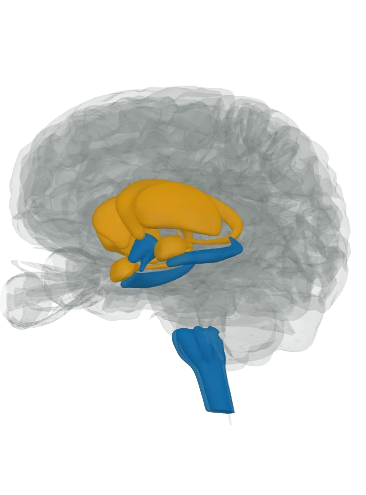
Made in: lit in blue
- Arcuate nucleus, lit as the hypothalamus: Its medial (inner) neurons make NPY together with AgRP and GABA and use it to raise appetite; ghrelin switches them on, and leptin, insulin and peptide YY hold them back. The source counts it among the places with the highest NPY levels. The nucleus has no shape of its own here, so the whole hypothalamus is lit. [2][1]
- C1 cell group, in the medulla oblongata: The C1 neurons of the ventrolateral medulla express several neuropeptides, NPY among them; the review names the lower brainstem, particularly the C1 group, as the main source of the NPY nerve endings in the paraventricular nucleus. The cells have no shape here, so the medulla is lit. [3][4]
- Hippocampus, lit as the hippocampus: High levels of NPY are made and act in the hypothalamus and the hippocampus. In the hippocampus it is made by GABA-releasing interneurons, local cells that connect neighbouring neurons; in the dentate gyrus, NPY released by interneurons supports the growth of new cells, and removing it cut that growth. [1][5]
Sent to: lit in orange
- Nucleus accumbens, lit as the caudate nucleus and putamen (reward and emotion circuits): Named with the amygdala as where NPY neurons interact with the dopamine reward pathways. It is not a separate shape here, so the caudate nucleus and putamen that hold it are lit. [1]
Also sent to, inside a part already lit in blue
- Paraventricular nucleus, inside the hypothalamus, already lit in blue (arcuate and brainstem → PVN): Its NPY nerve endings come from three sources: about 42% from adrenaline-making neurons (mainly C1, plus C3), 21% from noradrenaline-making neurons and the remaining 36% from the AgRP neurons of the arcuate nucleus. NPY released there with GABA by the arcuate endings appears to raise food intake. [3][1]
- Lateral hypothalamus, inside the hypothalamus, already lit in blue (arcuate → lateral hypothalamus): The arcuate’s NPY/AgRP/GABA neurons, in the most ventromedial part of the nucleus, project strongly to the lateral hypothalamus as well as to the paraventricular nucleus. [2]
- Also reached but inside the part already lit in blue: the arcuate’s NPY neurons directly contact and hold back their POMC/CART neighbours, and NPY regulates those cells’ release of beta-endorphin. [2]
- Not lit: the body’s sympathetic nerves, outside the brain. In the autonomic nervous system NPY is made mainly by sympathetic neurons, and there it is a strong constrictor of blood vessels. [1]
The pathways, as the sources name them
- Arcuate and brainstem → PVN, from the arcuate nucleus and the brainstem’s C1 and C3 cell groups to the paraventricular nucleus of the hypothalamus. NPY or noradrenaline put into the paraventricular nucleus raises feeding, and NPY there selectively raises carbohydrate intake; NPY placed there also releases corticotropin-releasing hormone, the first step of the stress-hormone chain. [3][1] See What part of the brain controls hunger?
- Arcuate → lateral hypothalamus, from the ventromedial part of the arcuate nucleus to the lateral hypothalamus. The same neurons are held back by leptin, insulin and peptide YY and switched on by ghrelin; when activated, they can produce ravenous eating. [2] See What part of the brain controls hunger?
- Reward and emotion circuits, from NPY neurons the source does not place, to the amygdala and the nucleus accumbens. The source calls NPY a calming (anxiolytic) peptide the body makes itself, whose levels can be changed by stress. [1] See What part of the brain controls the stress response?
Good to know
- NPY is a peptide 36 amino acids long; it was first isolated in 1982, from pig (porcine) hypothalamus. [1]
- It acts through its own receptors, mainly Y1, Y2, Y4 and Y6; Y1 and Y5 are linked to raising food intake, Y2 and Y4 to holding appetite back. [1] See What part of the brain controls hunger?
- All known NPY receptors are coupled to Gi/o proteins, so NPY is assumed to inhibit the cells it reaches; any excitatory effect of NPY is thought to come through disinhibition. [3]
- The roles the source lists include increasing food intake and the storage of energy as fat, reducing pain perception and affecting the daily (circadian) rhythm. [1]
- In the arcuate nucleus NPY also takes part in the growth-hormone system: GHRH neurons help control growth hormone release together with somatostatin and NPY. [2] See What part of the brain controls hormones?
What it takes part in
Each lit with every part that takes part: What part of the brain controls hunger? · What part of the brain controls the stress response? · What part of the brain controls hormones?
Where it is made, each on its own page: Arcuate nucleus; C1 cell group, in the medulla oblongata; Hippocampus.