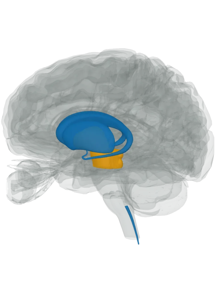
Made in: lit in blue
- Striatum, lit as the caudate nucleus and putamen: Its medium spiny neurons come in two main kinds; the direct-pathway (D1-type) ones express substance P, together with dynorphin, D1 dopamine receptors and adenosine A1 receptors, while the indirect-pathway ones use enkephalin instead. The striatum is the caudate nucleus and the putamen, so both are lit; the neurons themselves are not shapes in this atlas. [1]
- Dorsal horn, in the spinal cord (central canal): Pain-sensing nerve cells (nociceptors) have their cell bodies outside the cord, in the dorsal root ganglia; their fibres end in the layers (laminae) of the dorsal horn, and some use substance P as their transmitter there. Laminae I and II, the outermost, take the fibres that sense pain, temperature and itch. The atlas draws the cord only as its central canal, cropped just below the medulla, so that thin line is what is lit. [3][4][2]
Sent to: lit in orange
- Substantia nigra, lit as the midbrain (direct pathway): Direct-pathway neurons project straight to its pars reticulata, one of the basal ganglia’s two output nuclei. It is not a separate shape here, so the midbrain is lit. [1]
- Globus pallidus (direct pathway): The same neurons project to its internal segment (GPi), the other output nucleus. The atlas has one shape for the globus pallidus, so both its segments are lit. [1]
Also sent to, inside a part already lit in blue
- Dorsal horn neurons, inside the spinal cord (central canal), already lit in blue (nociceptor → dorsal horn): Different fibre types end in different layers of the dorsal horn and pass their signal on there, using glutamate or substance P; the fibres that respond to pain carry the two together. Most neurons of the outermost layers are interneurons, and the second-order neurons there send the signal on up to the brain. [3][2][4]
- Not lit: substance P and its NK1 receptor are widely spread through the brain, including regions that regulate emotion such as the hypothalamus, the amygdala and the periaqueductal grey; the cited sources do not name the cells that make it there, so nothing more is lit. [2]
- Not lit: the skin, muscles and joints, outside the brain. The same sensory fibres also release substance P from their outer endings there, where it widens blood vessels. [2]
The pathways, as the sources name them
- Direct pathway, from the striatum’s D1-type medium spiny neurons straight to the substantia nigra pars reticulata and the internal globus pallidus. It takes excitatory input from the cortex and the thalamus. A burst of activity in its inhibitory neurons briefly lifts the output nuclei’s brake on the thalamus; classic models link this to starting a movement, newer ones to favouring the wanted movement. [1] See What part of the brain controls movement?
- Nociceptor → dorsal horn, from pain-sensing nerve fibres, whose cell bodies lie in the dorsal root ganglia beside the cord, into the dorsal horn. The dorsal horn processes and integrates the sensory information arriving from the body; it takes input from these primary fibres and from systems that come down from the brain. [4][3] See What part of the brain feels touch, temperature and pain?
Good to know
- Substance P is an undecapeptide, a chain of 11 amino acids, cut from a larger precursor made by the preprotachykinin A gene (TAC1). [2]
- Its own receptor is the neurokinin 1 receptor (NK1R), found in both the central and the peripheral nervous system; the binding of substance P to it is associated with the transmission of pain signals. [2][6]
- Two messengers in one cell: the striatum’s medium spiny neurons are inhibitory cells that use GABA, and in the dorsal horn substance P comes out with the fast excitatory messenger glutamate. [1][2]
- The brain’s own counterweight: beta-endorphin acts in the spinal cord to cut the release of substance P, so less of the pain signal is passed up to the brain. [7]
- Discovery: Ulf von Euler and John H. Gaddum found it in 1931 as a tissue extract that made the gut contract; the “P” stands for “preparation” or “powder”. [2]
What it takes part in
Each lit with every part that takes part: What part of the brain feels touch, temperature and pain? · What part of the brain controls movement?
Where it is made, each on its own page: Striatum; Dorsal horn, in the spinal cord (central canal).