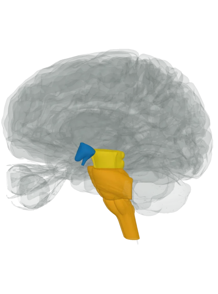
Side by side
| Compared | Dopamine | Serotonin |
|---|---|---|
| Where it is | Mainly the midbrain: the substantia nigra, a band on each side whose compact part (pars compacta) holds the dopamine neurons, and the ventral tegmental area on the floor of the midbrain near the midline. A smaller group sits in the arcuate and periventricular nuclei of the hypothalamus. About 400,000 dopamine neurons in all. [1][2][7] | The raphe nuclei, a ridge of cell groups along the midline of the brainstem: the dorsal and median raphe nuclei in the midbrain, the raphe pontis in the pons, and the raphe magnus, pallidus and obscurus in the medulla oblongata. Many of their neurons, but not most, make serotonin. [4][3] |
| What it does | Its four main pathways are involved in movement and motivation (nigrostriatal), the wanting of a reward and reinforcement learning (mesolimbic), cognition and executive functions (mesocortical), and controlling the pituitary gland’s release of some hormones, including prolactin (tuberoinfundibular). [2][1] | Serotonin from the raphe nuclei mainly promotes wakefulness and holds back rapid eye movement (REM) sleep; the lower raphe groups’ fibres to the spinal cord help control the release of enkephalins, which damp pain signals. [4][3] |
| Connects with | A few named routes to particular targets: the caudate nucleus and putamen (nigrostriatal), the nucleus accumbens (mesolimbic), the prefrontal cortex (mesocortical) and the pituitary gland (tuberoinfundibular). [2] | The upper (rostral) groups send fibres up across the forebrain, including the cerebral cortex, striatum, amygdala, hippocampus, thalamus and hypothalamus; the lower (caudal) groups send them down to the cerebellum and the spinal cord. [3][4][8] |
| Size measured from the model | Hypothalamus: 18 mm front to back, 20 mm top to bottom, 17 mm side to side [6] | Pons and medulla oblongata: 46 mm front to back, 66 mm top to bottom, 42 mm side to side [6] |
Size is the box each part fits in on this one model (hypothalamus; pons and medulla oblongata), not its volume. Real brains vary.
How far apart are they?
Measured from the model [6], from the hypothalamus to the pons and medulla oblongata:
- Centre to centre: 45 mm in a straight line.
- The centre of the pons and medulla oblongata is 26 mm further back, 37 mm lower and the same distance from the midline than the centre of the hypothalamus.
- Nearest points of the two shapes: about 7 mm apart.
A “centre” here is the average position of the points on the part’s 3D surface, so for a curved part such as a C shape it can sit outside the part itself. The nearest-points figure is measured between the corners of the simplified meshes, so a gap of 2 or 3 mm or less is within the model’s coarseness: read it as “touching or nearly so”.
How to tell them apart
- Where on the brainstem: dopamine’s main groups stop at the midbrain, while serotonin’s raphe nuclei run the whole length of the brainstem down to the medulla oblongata. Dopamine also has a source above the brainstem, in the hypothalamus; serotonin’s principal source, the raphe nuclei, lies wholly within the brainstem. [2][4]
- How it spreads: dopamine travels along a handful of named pathways to particular targets, while the axons of the upper raphe nuclei spread out across the entire brain, to many cortical and subcortical structures. [2][4]
- Where they meet: both reach the striatum. The caudate nucleus and putamen get dopamine from the substantia nigra and serotonin from the dorsal raphe nucleus. [2][8]
- What each is made from: dopamine from the amino acid tyrosine, by way of L-DOPA; serotonin from the amino acid tryptophan, by way of 5-hydroxytryptophan (5-HTP). [1][3]
- Mostly a gut chemical: about 90% of the body’s serotonin is made in the gut and only 1–2% is in the central nervous system. Dopamine in the blood cannot cross the blood–brain barrier, so the brain’s dopamine is made inside the brain. [3][1]