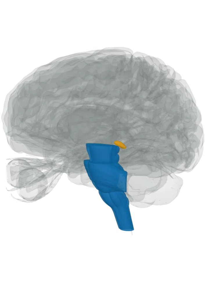
Side by side
| Compared | Serotonin | Melatonin |
|---|---|---|
| Where it is | The raphe nuclei, a ridge of cell groups along the midline of the brainstem: the dorsal and median raphe nuclei in the midbrain, the raphe pontis in the pons, and the raphe magnus, pallidus and obscurus in the medulla oblongata. Many of their neurons, but not most, make serotonin. [2][1] | One gland on the midline near the centre of the brain, between the two halves of the thalamus, just behind the third ventricle, resting between the superior colliculi of the midbrain. It is part of the epithalamus. [5][3] |
| What it does | Serotonin from the raphe nuclei mainly promotes wakefulness and holds back rapid eye movement (REM) sleep; the lower raphe groups’ fibres to the spinal cord help control the release of enkephalins, which damp pain signals. [2][1] | Light reduces melatonin production and darkness increases it, so blood levels fall in daylight and rise in the evening; melatonin helps set sleep patterns. After crossing time zones, its output can take several days to fit the new light-dark pattern. [3][5] |
| Connects with | The upper (rostral) groups send fibres up across the forebrain, including the cerebral cortex, striatum, amygdala, hippocampus, thalamus and hypothalamus; the lower (caudal) groups send them down to the cerebellum and the spinal cord. [1][2][6] | Light reaches it indirectly: light-sensitive ganglion cells in the retina signal the suprachiasmatic nucleus of the hypothalamus, which relays through the spinal cord and the superior cervical ganglion to the pineal gland. [5][3] |
| Size measured from the model | 46 mm front to back, 81 mm top to bottom, 42 mm side to side [4] | 11 mm front to back, 8 mm top to bottom, 6 mm side to side [4] |
Size is the box each part fits in on this one model (serotonin; melatonin), not its volume. Real brains vary.
How far apart are they?
Measured from the model [4], from the serotonin to the melatonin:
- Centre to centre: 30 mm in a straight line.
- The centre of the melatonin is 11 mm further back, 28 mm higher and the same distance from the midline than the centre of the serotonin.
- Nearest points of the two shapes: about 1 mm apart.
A “centre” here is the average position of the points on the part’s 3D surface, so for a curved part such as a C shape it can sit outside the part itself. The nearest-points figure is measured between the corners of the simplified meshes, so a gap of 2 or 3 mm or less is within the model’s coarseness: read it as “touching or nearly so”.
How to tell them apart
- One is made from the other: in the pineal gland, the amino acid tryptophan becomes 5-hydroxytryptophan (5-HTP), then serotonin, then N-acetylserotonin, then melatonin. [7][3]
- Messenger or hormone: serotonin acts between neurons, carried by raphe axons spread across the whole brain; melatonin is a hormone secreted by a gland that sits outside the blood–brain barrier. [2][3][5]
- Day and night: serotonin from the raphe nuclei mainly promotes wakefulness, while melatonin is produced in darkness, usually at night. [2][7]
- Where on the model: the raphe nuclei lie in the brainstem below and behind, the pineal gland sits just above the midbrain, between the superior colliculi, so the orange gland rests on top of the blue brainstem. [2][5]