Where is the medial forebrain bundle, and what does it join?
The medial forebrain bundle (MFB) is a bundle of nerve fibres running lengthwise along the base of the brain: from the midbrain, where the ventral tegmental area lies, forward through the lateral hypothalamus to the basal forebrain. It is part of the reward system, and carries fibres both ways. [1][2]
A fibre bundle has no shape of its own in this model; its two ends and the part it runs through are lit. [3]
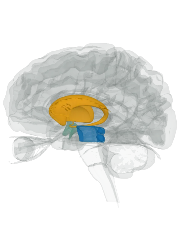
The brain opens cut down the midline and seen from the right, so the bundle's route along the base of the brain shows: the midbrain at the back, the hypothalamus in the middle and the basal forebrain in front. One end is lit in blue (the midbrain), the other in orange (the septum (septum of telencephalon), the caudate nucleus and the putamen), and the part it runs through in green (the hypothalamus); the bundle runs between them. Drag to turn, pinch or scroll to zoom; tap a lit part to name it.
The bundle itself is not drawn: BodyParts3D has no shape for any fibre bundle, so the parts at its two ends and the part it runs through are lit whole, on both sides. The ventral tegmental area is a small part of the blue midbrain, the lateral hypothalamus the outer zone of the green hypothalamus, and the nucleus accumbens the lowest front of the orange caudate nucleus and putamen; its branches into the frontal lobe are not lit. [3][1][4]
The two ends and the part it runs through: which colour is which
Back end: the midbrain, lit in blue: the midbrain, on both sides of the brain.
Its back end is in the midbrain, where the ventral tegmental area lies: the bundle carries fibres from there, and the mesolimbic pathway, the dopamine route from the ventral tegmental area to the nucleus accumbens, is one of its component pathways. [1][2]
Its front end is in the basal forebrain: it gathers fibres from the septal nuclei and the basal olfactory regions, and reaches the nucleus accumbens and the ventral striatum, which this atlas shows as the lowest front of the caudate nucleus and putamen. [1][4]
On the way: the lateral hypothalamus, lit in green: the hypothalamus, on both sides of the brain.
Between its two ends it passes, front to back, through the lateral hypothalamus, the outer zone of each half of the hypothalamus. That zone is not a separate shape in this atlas, so the whole hypothalamus is lit. [1][4]
The bundle runs between the blue and the orange parts, through the green one; its fibres are not drawn.
More about the medial forebrain bundle
Other names: MFB, fasciculus medialis telencephali [1]
It carries fibres in both directions, ascending and descending, and is one of the two major pathways joining the limbic forebrain, the midbrain and the hindbrain; the other is the dorsal diencephalic conduction system. [1]
It is the main tract for the ascending dopamine fibres. The mesolimbic pathway it carries ends in the ventral striatum, which includes the nucleus accumbens and the olfactory tubercle. [5][2]
The classic rat experiments: in 1953, at McGill University, James Olds and Peter Milner found that rats kept returning to the spot where they had received brief electrical stimulation in the septal area, and could be trained to press a lever for it. [5][6]
In later rat work, sites along the bundle gave the strongest reward effects; at the level of the lateral hypothalamus, rats pressed a lever several thousand times an hour, for days. [5]
In the human brain, a 2018 diffusion-imaging study of 55 healthy adults traced a superolateral branch from the ventral tegmental area through the anterior limb of the internal capsule to the nucleus accumbens and on to the frontal lobe, most of its connections reaching the superior frontal gyrus, the middle frontal gyrus and the lateral orbitofrontal region. [4]
That study keeps the older split into two parts: an inferomedial part, the evolutionarily older one, running through the lateral hypothalamus, and the younger superolateral branch, which connects to the cortex. [4]
Facts describe anatomy and normal function only. The 3D model is the adult male BodyParts3D brain (credits and licence); it has no shape for any fibre bundle, so a bundle is shown by the two parts it joins and, here, the part it runs through.