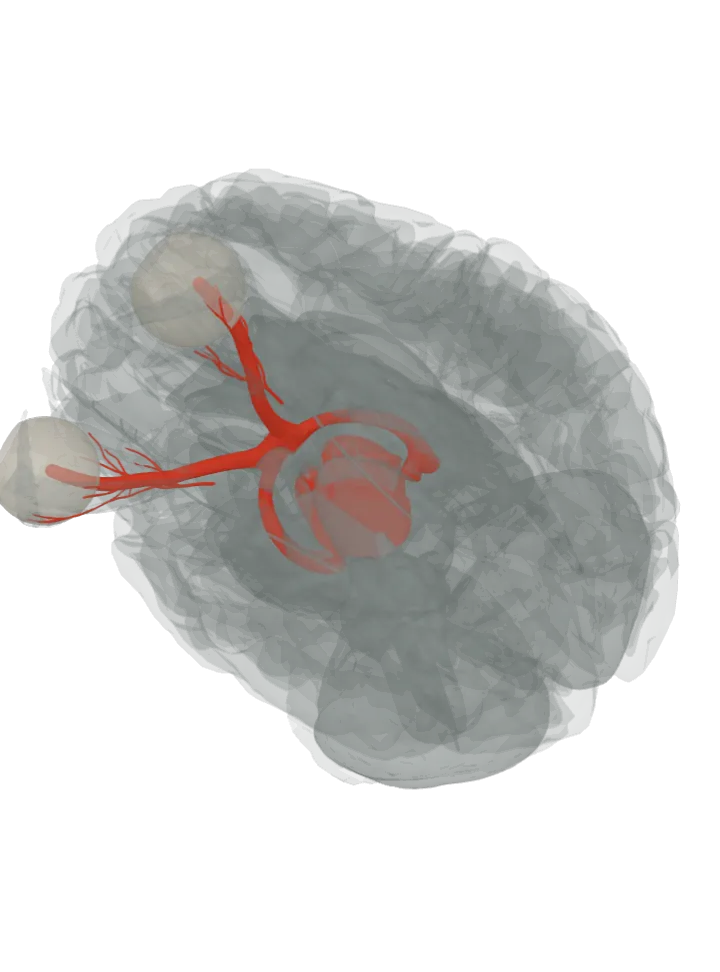
Light on one eye narrows both pupils: the signal runs along the optic nerve and tract to the pretectal area of the midbrain, on to the Edinger–Westphal nuclei of both sides, and back out in the oculomotor nerves to the iris sphincter of each eye. [1][2]
The atlas draws the optic nerve, chiasm, tract and oculomotor nerve but not the pretectal area; lit: the parts it runs through, in order.
The brain starts seen from below and a little from the left, where the optic nerves, the chiasm, the optic tracts and the oculomotor nerves lie on its underside. Step through lights one step at a time, about 1.2 s each, from the optic nerve to the iris; tap a step below to light it. The lit step is in the bright highlight colour, the steps before it stay lit in a paler tint of it and the steps still ahead are grey. Drag to turn, pinch or scroll to zoom. Step through is off because your device asks for reduced motion; tap each step instead.
The pretectal area, the Edinger–Westphal nucleus, the ciliary ganglion and the iris are not drawn: the midbrain is lit for the two nuclei, and the eyeball, drawn as its white outer coat, lights in its own colour with the last step. [3][1]
The route, in 6 steps
Light on the retina excites its ganglion cells, among them the photosensitive ganglion cells that report how bright it is. Their axons leave the eye in the optic nerve; some of them serve not sight but the pupil, bound for the pretectal area of the upper midbrain. [1][2]Where is the optic nerve?
At the optic chiasm the fibres from the inner (nasal) half of each retina cross, so each optic tract carries signals from both eyes. Most of the tract ends in the lateral geniculate body, on the way to conscious sight; the pupil fibres go on to the pretectal area, which hears from both retinas through the optic tract. [4][5][2]Where is the optic chiasm?Where is the optic tract?
The olivary pretectal nucleus lies just in front of the superior colliculus, at the top of the midbrain, and fires faster the brighter the light. Not a separate shape in this atlas; lit: the part that holds it, the midbrain. [2]Where is the midbrain?
Lights nothing new: the Edinger–Westphal nucleus sits in the same midbrain, beside the cerebral aqueduct at the level of the superior colliculus, and has no shape of its own here. Each pretectal nucleus signals the Edinger–Westphal nuclei of both sides, which is why light in one eye reaches both pupils. [6][1]
The Edinger–Westphal nucleus sends preganglionic parasympathetic fibres out with the oculomotor nerve, the nerve that also moves four of the six eye muscles. They relay in the ciliary ganglion behind the eye, which this atlas has no shape for. [1][6][7]Where is the oculomotor nerve?
Short ciliary nerves from the ciliary ganglion reach the iris sphincter, the ring of muscle round the pupil, and it tightens. Light in one eye narrows both pupils at once: the direct response in the lit eye and the consensual response in the other. [1]Where is the eyeball?
Lit with it: both eyeballs, in their own colour, drawn as the sclera, the white outer coat; the iris inside the front of each eye is not a separate shape. [3][1]
More about the pupillary light reflex
Other names: pupillary reflex, pupil light reflex, direct and consensual light reflex [1]
The pupils narrow about three times faster than they widen again when the light fades. [1]
It is not the route you see with: conscious sight runs from the optic tract to the lateral geniculate body and on to the occipital lobe, while this reflex turns off to the midbrain before the thalamus. [8][2]
The same Edinger–Westphal nucleus also works the ciliary muscle, which changes the shape of the lens to focus on near things (accommodation). [6]
Facts describe anatomy and normal function only. The 3D model is the adult male BodyParts3D brain (credits and licence); it has no shape for tracts, so a pathway is shown by the parts it runs through.