It stays on the same side (ipsilateral) and never reaches conscious awareness: it carries body position from the trunk and legs to the cerebellum of the same side, through the inferior and superior cerebellar peduncles, never passing through the thalamus or reaching the cortex. [1][2]
A tract has no shape of its own in this atlas; lit: the parts it runs through, in order.
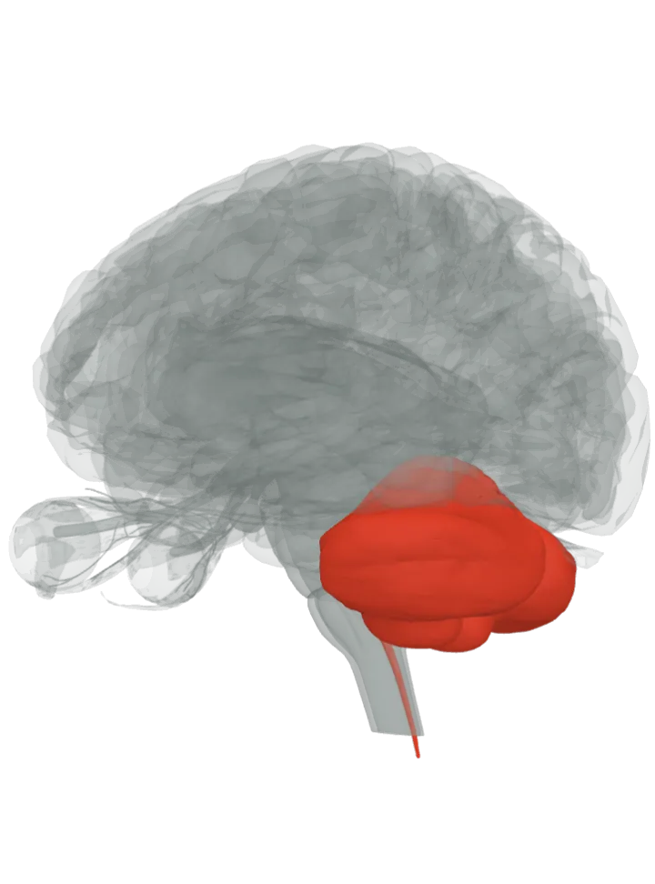
The brain starts seen from the left, a little from behind and below, so the spinal cord, the back of the brainstem and the cerebellum it climbs into all show. Step through lights one step at a time, about 1.2 s each, bottom to top; tap a step below to light it. The lit step is in the bright highlight colour, the steps before it stay lit in a paler tint of it and the steps still ahead are grey. Drag to turn, pinch or scroll to zoom. Step through is off because your device asks for reduced motion; tap each step instead.
Each step lights the whole part. The atlas draws the spinal cord only as its central canal and has no shape for Clarke's nucleus, the tracts or the peduncles, so the peduncle step lights, as the cerebellar peduncles page does, the cerebellum and the midbrain, pons and medulla oblongata the three pairs join, though this route uses only the inferior and superior pair. [3][2]
The route, in 3 steps
Position signals from the muscles, tendons and joints of the trunk and legs enter the spinal cord through the dorsal roots. For the dorsal spinocerebellar tract they synapse in Clarke's nucleus (the posterior thoracic nucleus), and the second-order axons climb the outer edge of the cord on the same side. The atlas draws the cord only as its central canal, so Clarke's nucleus and the tracts are not drawn. [1]Where is the spinal cord?
Crossing twice: the ventral spinocerebellar tract crosses to the opposite side in the spinal cord and crosses back inside the cerebellum, so it too ends on the side it started from. The atlas has no shape for either crossing, so neither is marked. [1]
The dorsal tract climbs into the medulla oblongata and turns back into the cerebellum through the inferior cerebellar peduncle of the same side. The ventral tract climbs higher, through the pons, and enters the cerebellum through the superior cerebellar peduncle, the stalk that joins it to the midbrain. The cerebellar peduncles have no shape in this atlas; lit: the cerebellum and the three brainstem parts they join. [1][2]Where are the cerebellar peduncles?
The fibres end in the cerebellar cortex on the side they started from; the ventral tract's in the anterior lobe and in the vermis of the posterior lobe. The cerebellum folds this running report of limb position in with its other inputs to fine-tune movement as it happens, and the signal is never felt. [1][4]Where is the cerebellum?
How it differs from the dorsal column–medial lemniscus pathway: both carry body position from the limbs, but the dorsal column pathway crosses in the medulla, relays in the thalamus and ends in the postcentral gyrus, where position is consciously felt; the spinocerebellar tracts stay on one side and end in the cerebellum, unfelt. [5][1]Where does the dorsal column–medial lemniscus pathway run?
Four tracts make up this unconscious route: the dorsal and ventral spinocerebellar tracts serve the trunk and legs, and the cuneocerebellar and rostral spinocerebellar tracts the arm. [1]
The cuneocerebellar tract, the arm and neck counterpart of the dorsal tract, starts in the accessory cuneate nucleus of the medulla and enters the inferior cerebellar peduncle of the same side. [1]
The rostral spinocerebellar tract carries signals from the Golgi tendon organs of the upper half of the body. [1]
Facts describe anatomy and normal function only. The 3D model is the adult male BodyParts3D brain (credits and licence); it has no shape for tracts, so a pathway is shown by the parts it runs through.