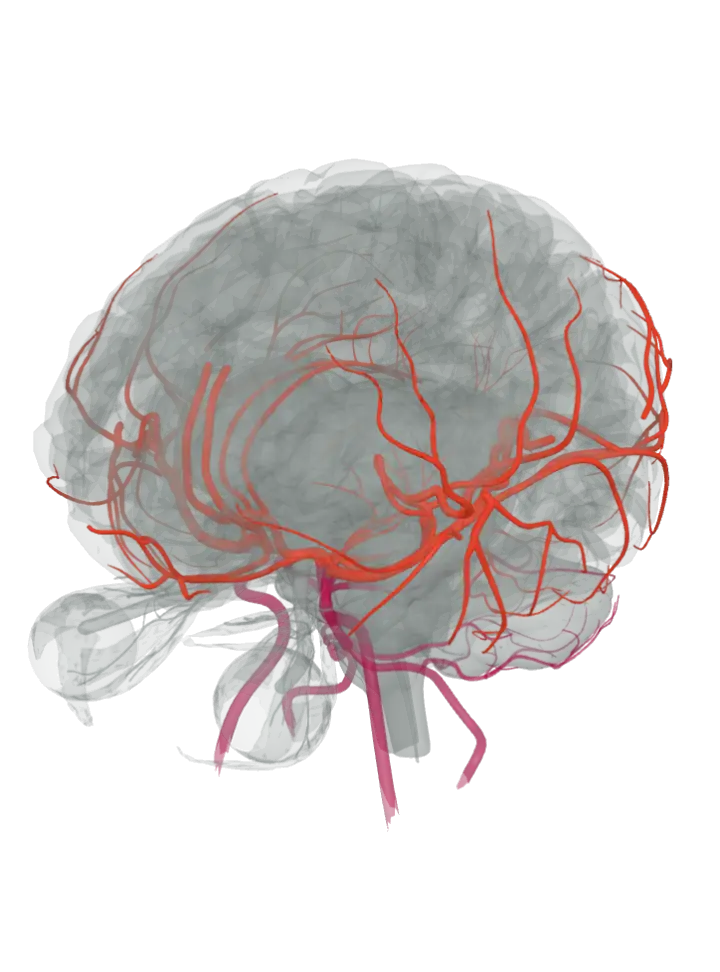
The front route: the internal carotid arteries
Each internal carotid artery leaves the common carotid in the neck, at about the level of the fourth neck vertebra, climbs into the skull through the carotid canal, passes beside the pituitary gland and ends under the brain by dividing into the anterior and middle cerebral arteries. On the way it gives off the ophthalmic artery to the eye and the posterior communicating artery. This front (anterior) route carries the blood for most of each cerebral hemisphere: the frontal and parietal lobes and most of the temporal lobe. [7][1]
Each lit on its own page: internal carotid artery, anterior cerebral artery and middle cerebral artery.
The back route: the vertebral and basilar arteries
The two vertebral arteries branch from the subclavian arteries at the base of the neck, rise through holes in the side of the upper six neck vertebrae, enter the skull through the foramen magnum and join at the lower edge of the pons to form the single basilar artery, which runs up the front of the pons. Each vertebral artery gives off a posterior inferior cerebellar artery, and the basilar artery the anterior inferior and superior cerebellar arteries, so this back (posterior) route feeds the brainstem and the cerebellum. At the top of the brainstem the basilar artery splits into the two posterior cerebral arteries, which carry the back route on to the occipital lobes and the underside of the temporal lobes. [8][9]
Each lit on its own page: vertebral artery, basilar artery, cerebellar arteries and posterior cerebral artery.
Where the two routes meet: the circle of Willis
Under the brain, around the optic chiasm and the pituitary stalk, the two routes are joined into a ring: the anterior communicating artery links the left and right anterior cerebral arteries, and on each side a posterior communicating artery links the internal carotid to the posterior cerebral artery. The ring's shape varies a lot from person to person; often one of its links is thin or missing. [2]
Each lit on its own page: circle of Willis, anterior communicating artery and posterior communicating artery.
How much blood the brain takes
An adult brain takes about 750 millilitres of blood a minute, roughly 15% of everything the heart pumps out. [1]
Every artery page, each lit on its own
- Circle of Willis A ring of linked arteries on the underside of the brain, in the hollow between the optic chiasm in front and the brainstem behind: the two internal carotids and the top of the basilar artery feed it.
- Middle cerebral artery The largest branch of the internal carotid: runs sideways under the brain, then up into the lateral (Sylvian) fissure over the insula, and fans out over the outer face of the hemisphere.
- Anterior cerebral artery Starts from the internal carotid under the front of the brain, runs forward above the optic nerve into the deep groove between the hemispheres, then arches back over the corpus callosum (as the pericallosal and callosomarginal arteries).
- Posterior cerebral artery The two end branches of the basilar artery, at the top of the brainstem: each curves back around the midbrain to the underside of the temporal lobe and the inner face of the occipital lobe.
- Basilar artery A single midline artery, about 3 cm long, lying in the groove on the front of the pons; formed where the two vertebral arteries join at the lower edge of the pons.
- Internal carotid artery Rises up each side of the neck, enters the skull through the carotid canal, runs through the cavernous sinus beside the pituitary and ends under the brain beside the optic chiasm; drawn here from the level of the foramen magnum up.
- Vertebral artery Climbs through the neck vertebrae, enters the skull through the foramen magnum and runs up the front of the medulla to join its partner at the lower edge of the pons; drawn here from the foramen magnum up (the part inside the skull).
- Anterior communicating artery A short cross-link, only a few millimetres long, between the left and right anterior cerebral arteries in front of the optic chiasm, at the front of the circle of Willis.
- Posterior communicating artery A short artery on each side of the circle of Willis, running back from the internal carotid to the posterior cerebral artery, beside the mammillary bodies and close to the oculomotor nerve.
- Cerebellar arteries Three arteries on each side, from top to bottom: the superior cerebellar from the top of the basilar, the anterior inferior cerebellar from its lower part, the posterior inferior cerebellar from the vertebral artery; each winds back around the brainstem to the cerebellum.