How to read it
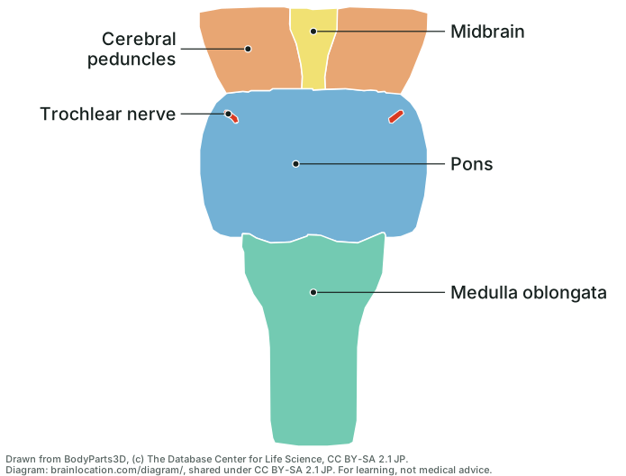
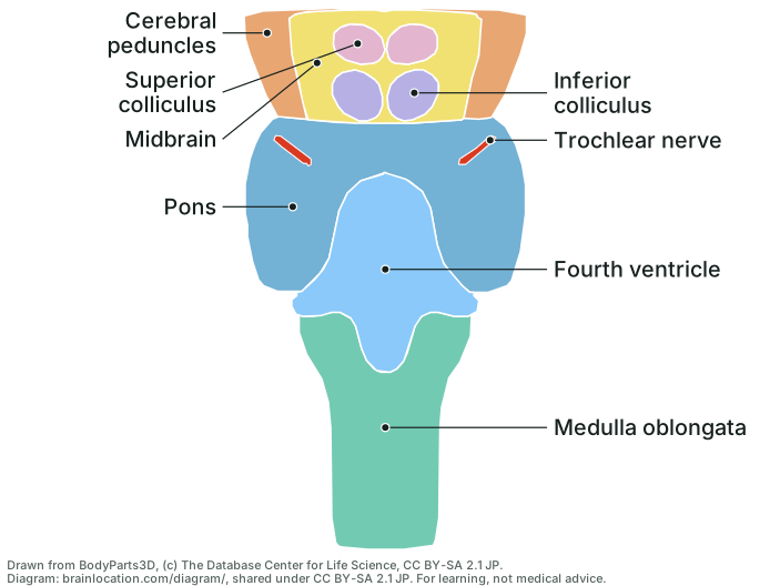
- Top to bottom the brainstem has three parts: the midbrain, the pons and the medulla oblongata, which carries on below as the spinal cord (Wikipedia: Brainstem; OpenStax A&P 2e, 13.2).
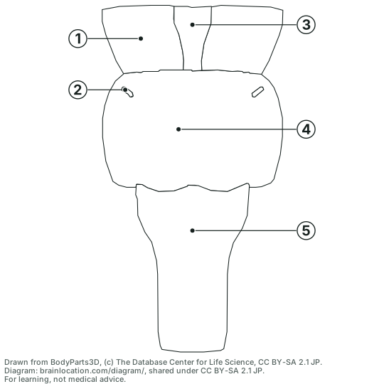
- From the front, the midbrain is mostly its two cerebral peduncles, the thick stalks of descending fibres that splay up and out towards each hemisphere; the yellow wedge between them is the floor of the gap they leave, the interpeduncular fossa, where the oculomotor nerve comes out (Wikipedia: Cerebral peduncle). Below, the pons is the widest part, a rounded bulge of fibres crossing to the cerebellum (Wikipedia: Pons).
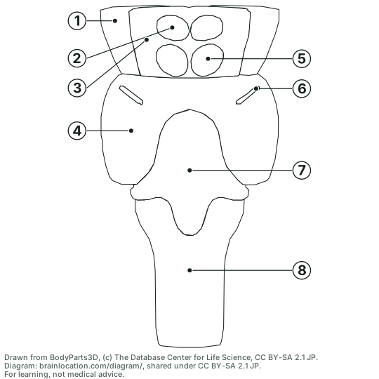
- From the back, the roof of the midbrain carries four bumps, two superior colliculi above two inferior colliculi (together the corpora quadrigemina): the superior pair turns the eyes and head toward things you see, hear or feel, the inferior pair is the midbrain’s hearing relay (Wikipedia: Superior colliculus; Wikipedia: Inferior colliculus). Below them the diamond-shaped fourth ventricle lies on the back of the pons and upper medulla; its floor is the rhomboid fossa (Wikipedia: Fourth ventricle; Wikipedia: Rhomboid fossa).
- Measured on the model: the brainstem (midbrain to the lowest point of the medulla) is about 81 mm tall; the pons is about 42 mm across and 29 mm tall, the midbrain about 17 mm tall, the medulla shape 41 mm long, and the fourth ventricle about 28 mm at its widest (BodyParts3D 4.0, © DBCLS, CC BY-SA 2.1 Japan). On the sheets every millimetre of the model is drawn as two units. Real brains differ.
Where each cranial nerve leaves the brainstem
Ten of the twelve cranial nerves (III to XII) attach to the brainstem; I and II attach to the forebrain (Wikipedia: Cranial nerves; Wikipedia: Brainstem). The model has a shape for only one of these roots, the trochlear nerve (IV), drawn on both sheets; the others are listed here, not drawn. All twelve are on the cranial nerves table.
- III, oculomotor: the front of the midbrain, between the two cerebral peduncles (the interpeduncular fossa) (Wikipedia: Cerebral peduncle; Wikipedia: Oculomotor nerve).
- IV, trochlear: the back of the midbrain just below the inferior colliculi, the only nerve to leave the back; it wraps forward round the peduncle (Wikipedia: Trochlear nerve).
- V, trigeminal: the side of the pons, about halfway down, a large sensory root with a small motor root beside it (Wikipedia: Trigeminal nerve).
- VI, abducens: the groove between the pons and the medulla, close to the midline, just above the pyramid (Wikipedia: Abducens nerve).
- VII, facial: the same groove further out, between VI and VIII (Wikipedia: Facial nerve).
- VIII, vestibulocochlear: the outer end of that groove, in the angle between pons, medulla and cerebellum (the cerebellopontine angle) (Wikipedia: Vestibulocochlear nerve).
- IX, glossopharyngeal: the side of the upper medulla, as rootlets in the groove behind the olive (Wikipedia: Glossopharyngeal nerve).
- X, vagus: the same groove behind the olive, as a row of rootlets just below IX (Wikipedia: Vagus nerve).
- XI, accessory: mostly the upper spinal cord: its rootlets climb into the skull beside the medulla (older accounts add a cranial root from the medulla) (Wikipedia: Accessory nerve).
- XII, hypoglossal: the front of the medulla, as rootlets in the groove between the pyramid and the olive (Wikipedia: Hypoglossal nerve).
How it is drawn
- The shapes are not traced by hand: the BodyParts3D meshes of the midbrain, cerebral peduncles, colliculi, pons, medulla oblongata, fourth ventricle and trochlear nerve, the same ones the 3D brain on this site shows, are projected straight onto the page (no perspective) on a grid of 0.125 mm cells, and each cell keeps the part nearest the eye. Everything else in the model (cerebrum, thalamus, cerebellum) is left off.
- Two parts are drawn through the surface on the back view. In the model the colliculi lie 1 to 3 mm under the outer surface of the midbrain shape, and the trochlear nerve runs under the back of the pons shape, so a strict view would hide them; they are painted over it where they lie. The trochlear nerve is cut off 22 mm in front of the model’s centre, just past the front of the peduncles: only its root and its turn round the brainstem are drawn, not its run forward to the eye socket.
- Each label starts at the point deepest inside its own shape:
test/diagram.test.cjschecks that every dot lies inside the outline drawn for that part and that the line of sight through the dot meets that part first (for the parts drawn through the surface: meets that part at all).
What it does not show
- The three pairs of cerebellar peduncles (superior to the midbrain, middle to the pons, inferior to the medulla) have no shapes of their own in the model, so they are not labelled; with the cerebellum off, the brainstem shapes simply end where they would leave (Wikipedia: Cerebellar peduncle; BodyParts3D 4.0, © DBCLS, CC BY-SA 2.1 Japan).
- The front of the medulla has two ridges either side of the midline, the pyramids, and an oval bulge beside each, the olive, over the inferior olive; the model’s medulla is one smooth shape, so neither is drawn (Wikipedia: Medullary pyramids; Wikipedia: Olivary body).
- The nerve roots other than IV (above), and the nuclei inside the brainstem such as the substantia nigra and red nucleus, which a surface view cannot show.
- It is one adult brain. The drawing is derived from BodyParts3D and carries its licence, CC BY-SA 2.1 Japan: copy, hand out and reuse it with the credit line. Standalone SVG files: front labelled and blank, back labelled and blank.