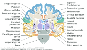
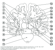
The brain cut from ear to ear through the thalamus, as on a coronal slide or scan: the face of the cut, computed from the 3D model this site uses. Below it, the same slice blank and numbered, with an answer key.
Coronal section: the brain cut from ear to ear through the thalamus
Coronal section through the thalamus about 4 mm in front of its middle, where the globus pallidus is cut too, seen from the front; the brain’s left half on the right of the sheet, as when you face someone. 23 parts labelled.
Drawn from BodyParts3D, (c) The Database Center for Life Science, CC BY-SA 2.1 JP. Diagram: brainlocation.com/diagram/, shared under CC BY-SA 2.1 JP (credits). For learning, not medical advice.
Coronal section, blank: name the 23 numbered parts
Coronal section through the thalamus about 4 mm in front of its middle, where the globus pallidus is cut too, seen from the front; the brain’s left half on the right of the sheet, as when you face someone.
Drawn from BodyParts3D, (c) The Database Center for Life Science, CC BY-SA 2.1 JP. Diagram: brainlocation.com/diagram/, shared under CC BY-SA 2.1 JP (credits). For learning, not medical advice.
Which cut this is
A coronal plane runs from ear to ear and splits the brain into a front and a back part; this sheet shows the face of the back part, looked at from the front. The other standard views are on the labelled diagram (side, midline, underside) and the horizontal section.
Work from the midline outwards: the third ventricle between the two thalami, then the internal capsule, a white band, between the thalamus and caudate nucleus on the inside and the putamen and globus pallidus on the outside, then the insula under the outer cortex.
The lateral ventricles sit right under the corpus callosum, with the body of the caudate nucleus in their outer wall. The fornix shows as a small patch under them, just above the third ventricle.
The hippocampus is low down on each side, on the inner edge of the temporal lobe, against the small lower (temporal) horn of the lateral ventricle, with the parahippocampal gyrus under it.
In this model the same vertical cut passes through the midbrain and the pons below the thalamus, so the brainstem shows in the middle at the bottom.
How it is drawn
The shapes are not traced by hand: the BodyParts3D meshes the 3D brain on this site shows are cut on one flat plane, on a grid of 0.25 mm cells. For each part the plane passes through, the half beyond the cut is removed and the rest is depth-buffered from the cut side, as the midline view is; a cell belongs to the part when the first surface of that part the eye meets is the inside of its far wall, which only happens when the cell lies within it. Where a cell is inside two parts (a nucleus within the white matter) the smaller, inner one wins.
So only parts the cut really passes through are drawn, and every label names one of them: test/diagram.test.cjs rebuilds the cut, and for each label checks that its dot lies inside the outline drawn for that part and inside the part's own 3D shape, tested on its own by casting rays from the dot in six directions.
The claustrum and the external capsule (the thin grey sheet and the white layers between the putamen and the insula) have no shape of their own in the model; they are part of the white matter on this sheet. In the model the amygdala lies further forward than this cut, so it is not on it.
Parts the atlas has no shape for at all (the substantia nigra and red nucleus inside the midbrain, the thalamic nuclei) are not drawn; their pages light the part that holds them. The tentorium, a fold of the brain’s outer covering, is in the model but left off: it is not brain.
It is one adult brain, with small gaps where the model’s shapes do not quite meet (left white). Real brains differ, and a slide or scan a few millimetres further forward or back shows the parts in other proportions, so learn the order of the parts rather than their exact shapes.
The drawing is derived from BodyParts3D and carries its licence, CC BY-SA 2.1 Japan: copy, hand out and reuse it with the credit line. Standalone SVG files: labelled, blank.