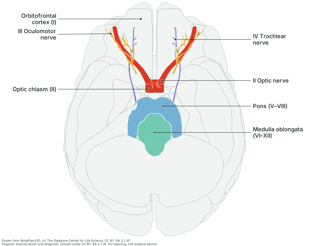
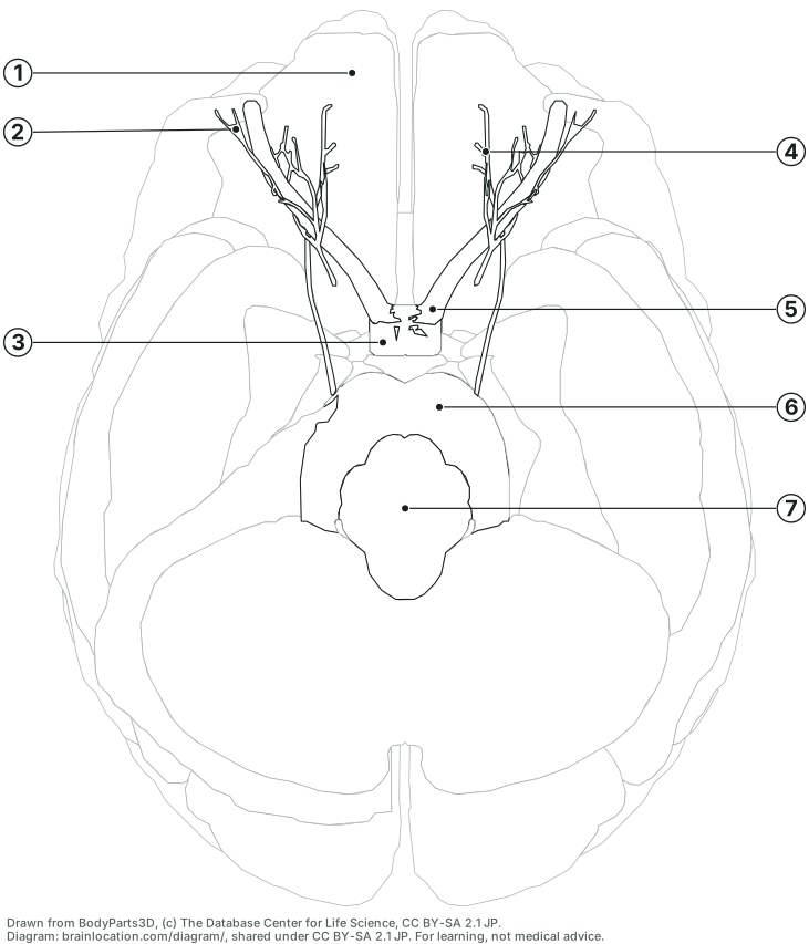
All twelve, I to XII, and which are drawn
The same rows, numerals and sources as the cranial nerves table. Each nerve is a pair, one on each side.
- I Olfactory nerve: Not a shape in the model; marked on Orbitofrontal cortex (I). Leaves: not from the brainstem: its fine fibres rise from the lining of the nose through the cribriform plate of the skull and end in the olfactory bulb, under the front of the brain. The bulb is not a shape in this atlas; labelled on the sheet: the orbitofrontal cortex it lies against. (Wikipedia: Olfactory nerve; Wikipedia: Olfactory bulb). Does: Smell. (Wikipedia: Olfactory nerve; OpenStax Anatomy and Physiology 2e, 13.4 The Peripheral Nervous System).
- II Optic nerve: Drawn (II Optic nerve, Optic chiasm (II)). Leaves: not from the brainstem: from the back of each eye it runs through the optic canal to the optic chiasm under the front of the brain, where about half its fibres cross; the optic tracts then carry on to the thalamus. (Wikipedia: Optic nerve; Wikipedia: Cranial nerves). Does: Vision: it carries everything the retina sees. (Wikipedia: Optic nerve; OpenStax Anatomy and Physiology 2e, 13.4 The Peripheral Nervous System).
- III Oculomotor nerve: Drawn (III Oculomotor nerve). Leaves: the front of the midbrain, in the hollow between the two cerebral peduncles (the interpeduncular fossa). (Wikipedia: Oculomotor nerve). Does: Works four of the six muscles that turn each eye (the superior, inferior and medial rectus and the inferior oblique), lifts the upper eyelid, and narrows the pupil and focuses the lens. (Wikipedia: Oculomotor nerve; OpenStax Anatomy and Physiology 2e, 13.4 The Peripheral Nervous System).
- IV Trochlear nerve: Drawn (IV Trochlear nerve). Leaves: the back of the midbrain, just below the inferior colliculi: the only cranial nerve to leave the back of the brainstem. It then winds forward round the midbrain. (Wikipedia: Trochlear nerve). Does: Works one eye muscle, the superior oblique, which turns the eye down and inward. (Wikipedia: Trochlear nerve; OpenStax Anatomy and Physiology 2e, 13.4 The Peripheral Nervous System).
- V Trigeminal nerve: Not a shape in the model; marked on Pons (V–VIII). Leaves: the side of the pons, as a large sensory root and a small motor root, where the pons joins the middle cerebellar peduncle. (Wikipedia: Trigeminal nerve). Does: Feeling (touch, pain, temperature) from the face, mouth and inside of the nose, and the muscles of chewing. (Wikipedia: Trigeminal nerve; OpenStax Anatomy and Physiology 2e, 13.4 The Peripheral Nervous System).
- VI Abducens nerve: Not a shape in the model; marked on Pons (V–VIII) and Medulla oblongata (VI–XII). Leaves: the border of the pons and the medulla (the pontomedullary junction), close to the midline, inward of the facial nerve. (Wikipedia: Abducens nerve; Wikipedia: Cranial nerves). Does: Works the lateral rectus, the muscle that turns each eye outward. (Wikipedia: Abducens nerve; OpenStax Anatomy and Physiology 2e, 13.4 The Peripheral Nervous System).
- VII Facial nerve: Not a shape in the model; marked on Pons (V–VIII) and Medulla oblongata (VI–XII). Leaves: the border of the pons and the medulla, further out to the side than the abducens, in the angle between the pons and the cerebellum (the cerebellopontine angle). (Wikipedia: Facial nerve; Wikipedia: Cranial nerves). Does: Works the muscles of facial expression, carries taste from the front two-thirds of the tongue, and drives the tear glands and two of the salivary glands (the submandibular and sublingual). (Wikipedia: Facial nerve; OpenStax Anatomy and Physiology 2e, 13.4 The Peripheral Nervous System).
- VIII Vestibulocochlear nerve: Not a shape in the model; marked on Pons (V–VIII) and Medulla oblongata (VI–XII). Leaves: the border of the pons and the medulla, just outside the facial nerve, in the cerebellopontine angle. (Wikipedia: Vestibulocochlear nerve; Wikipedia: Cranial nerves). Does: Hearing, from the cochlea, and balance: the sense of head position and movement from the vestibular organs of the inner ear. (Wikipedia: Vestibulocochlear nerve; OpenStax Anatomy and Physiology 2e, 13.4 The Peripheral Nervous System).
- IX Glossopharyngeal nerve: Not a shape in the model; marked on Medulla oblongata (VI–XII). Leaves: the side of the upper medulla, as rootlets in the groove behind the olive, just above the vagus nerve. (Wikipedia: Glossopharyngeal nerve). Does: Taste and touch from the back third of the tongue and the throat, one throat muscle used in swallowing (the stylopharyngeus), the parotid salivary gland, and signals on blood pressure and blood oxygen from the carotid sinus and carotid body. (Wikipedia: Glossopharyngeal nerve; OpenStax Anatomy and Physiology 2e, 13.4 The Peripheral Nervous System).
- X Vagus nerve: Not a shape in the model; marked on Medulla oblongata (VI–XII). Leaves: the side of the medulla, as a row of rootlets in the groove behind the olive, below the glossopharyngeal nerve. (Wikipedia: Vagus nerve). Does: The parasympathetic supply to the heart, lungs and most of the gut, the muscles of the throat and voice box, and feeling from the organs of the chest and abdomen. (Wikipedia: Vagus nerve; OpenStax Anatomy and Physiology 2e, 13.4 The Peripheral Nervous System).
- XI Accessory nerve: Not a shape in the model; marked on Medulla oblongata (VI–XII). Leaves: mostly the upper spinal cord: rootlets from about its top five or six segments join and rise through the foramen magnum into the skull. A small cranial root from the side of the medulla is often counted as part of the vagus instead. (Wikipedia: Accessory nerve; Wikipedia: Cranial nerves). Does: Works two neck and shoulder muscles: the sternocleidomastoid, which turns and tilts the head, and the trapezius, which shrugs the shoulder. (Wikipedia: Accessory nerve; OpenStax Anatomy and Physiology 2e, 13.4 The Peripheral Nervous System).
- XII Hypoglossal nerve: Not a shape in the model; marked on Medulla oblongata (VI–XII). Leaves: the front of the medulla, as rootlets in the groove between the pyramid and the olive. (Wikipedia: Hypoglossal nerve). Does: Works the muscles of the tongue (all but one, the palatoglossus), for speaking, chewing and swallowing. (Wikipedia: Hypoglossal nerve; OpenStax Anatomy and Physiology 2e, 13.4 The Peripheral Nervous System).
How to read it
- You are looking up at the brain from below, front at the top. As on any view from below, the brain’s left half is on the right of the sheet. The eyeballs and eye muscles are left off, so the nerves that run forward into the eye sockets end in mid-air at the top.
- The two optic nerves (II) run back from the eye sockets and meet at the X-shaped optic chiasm, just in front of where the pituitary gland hangs; behind it the optic tracts, drawn pale, curve round the brainstem (Wikipedia: Optic nerve; Wikipedia: Cranial nerves). In this model each optic nerve spans about 49 mm from front to back and the chiasm about 15 mm from side to side (BodyParts3D 4.0, © DBCLS, CC BY-SA 2.1 Japan).
- The oculomotor nerve (III) is modelled only inside the eye socket, where it splits into the branches that reach four of the six eye muscles; its root at the front of the midbrain, between the cerebral peduncles, is not a shape, so the stretch from there to the socket is not drawn (Wikipedia: Oculomotor nerve; BodyParts3D 4.0, © DBCLS, CC BY-SA 2.1 Japan).
- The thin trochlear nerve (IV) leaves the back of the midbrain and winds forward round its side; on the sheet it runs from beside the brainstem to the eye socket, about 79 mm from front to back in this model, the longest course inside the skull of any cranial nerve (Wikipedia: Trochlear nerve; BodyParts3D 4.0, © DBCLS, CC BY-SA 2.1 Japan).
- Ten of the twelve leave the brainstem; I and II join the forebrain instead (Wikipedia: Cranial nerves). The pons and medulla are coloured as on the cranial nerves table, so the numerals in brackets read as: these nerves leave from this part. VI, VII and VIII leave from the groove where the pons meets the medulla, which is why they are listed on both.
How it is drawn
- The same projection as the underside view, at 1.5 times its scale: every BodyParts3D mesh, the ones the 3D brain on this site shows, is projected straight up onto the page (no perspective) on a grid of cells, each keeping the part nearest the eye. Nothing is traced by hand.
- What is coloured and every numeral are read from the data the cranial nerves table is built from, never typed here: the coloured parts are the ones that page’s 3D view shows before you press a row, and a label’s numeral is the row the nerve has there.
test/diagram.test.cjsreads the built table and fails if any numeral on this sheet differs from its row. - The nerve shapes are drawn through anything in front of them: where the part nearest the eye is pale and a nerve lies behind it, the cell is given to the nerve, at its true projected place. Each label starts at the point deepest inside its own shape; the test checks every dot lies inside the outline drawn for its part and inside that part’s projected extent in the model.
What it does not show
- Nine of the twelve nerves: I and V–XII have no shape in the model (BodyParts3D 4.0, © DBCLS, CC BY-SA 2.1 Japan), so none is drawn as a line; each is marked by numeral on the part it leaves from (the olfactory bulb of I lies under the orbitofrontal cortex, next to the midline; Wikipedia: Olfactory nerve; Wikipedia: Olfactory bulb).
- Hidden from below, so not labelled: the midbrain, which from below lies behind the cerebral peduncles and the pons, so it is coloured but out of sight; and the spinal cord, which this model has only as its central canal, a thread about 2 mm wide inside the lowest part of the medulla.
- The eyeballs and eye muscles, which the model holds as context around the nerves, are left off. The pituitary gland is not drawn either: it hangs just below and behind the chiasm, and the chiasm is drawn through it. One adult brain: real nerves differ in length and course. The drawing is derived from BodyParts3D and carries its licence, CC BY-SA 2.1 Japan: copy, hand out and reuse it with the credit line. Standalone SVG files: labelled and blank.