The 10 parts, and what each does
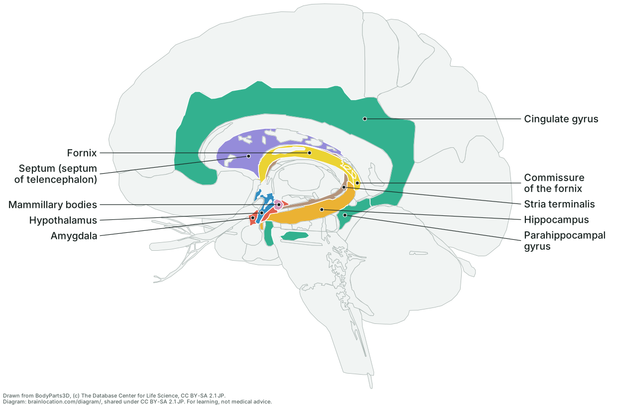
- Hippocampus: In the inner temporal lobe; it is central to forming new memories of events and facts and to spatial memory. (Wikipedia: Hippocampus).
- Amygdala: An almond-shaped cluster in front of the hippocampus; it processes emotion, fear above all, and tags memories with emotion. (Wikipedia: Amygdala).
- Cingulate gyrus: The fold arching over the corpus callosum; it takes part in emotion, learning and memory. (Wikipedia: Cingulate cortex).
- Parahippocampal gyrus: The fold just below the hippocampus; it takes part in memory encoding and retrieval and in recognising scenes and places. (Wikipedia: Parahippocampal gyrus).
- Hypothalamus: At the base, below the thalamus; it links the limbic system to the body, through hormones and drives such as hunger, thirst and body temperature. (Wikipedia: Hypothalamus; Wikipedia: Limbic system).
- Fornix: The C-shaped fibre bundle that arches from each hippocampus up under the corpus callosum and down to the mammillary bodies; it is the main output of the hippocampus and the second link of the Papez circuit. (Wikipedia: Fornix (neuroanatomy); Wikipedia: Papez circuit).
- Commissure of the fornix: Where the bodies of the two fornices meet, under the back of the corpus callosum: a thin triangular sheet of fibres joining the left and right hippocampus across the midline. (Wikipedia: Hippocampal commissure; Wikipedia: Fornix (neuroanatomy)).
- Mammillary bodies: Two small round bumps at the back of the hypothalamus where the fornix ends; they pass memory signals on to the anterior thalamic nuclei through the mammillothalamic tract. (Wikipedia: Mammillary body; Wikipedia: Papez circuit).
- Septum (septum of telencephalon): On the midline under the front of the corpus callosum: the septal area, which holds the septal nuclei the cited source lists among the limbic system’s subcortical parts. The atlas names this one shape “septum of telencephalon”, so it is not labelled the septum pellucidum here. (Wikipedia: Limbic system; Wikipedia: Septum pellucidum; BodyParts3D 4.0 (3D model measured on this page), © DBCLS, CC BY-SA 2.1 Japan).
- Stria terminalis: A thin fibre band along the floor of each lateral ventricle, between the thalamus and the caudate nucleus: a major output path of the amygdala, to the septal nuclei, the hypothalamus and the thalamus. (Wikipedia: Stria terminalis; Wikipedia: Limbic system).
Honest limit: it is no longer seen as one unit responsible for emotion, and which structures belong to it is debated, so read the lit set as the parts most often named, not a closed list. (Wikipedia: Limbic system).
How to read it
- Front of the brain is on the left. The green arch over the pale corpus callosum is the cingulate gyrus; it carries on round the back and down into the parahippocampal gyrus on the inner face of the temporal lobe. Together they are the ring Broca named the limbic lobe, from the Latin limbus, edge (Wikipedia: Limbic system; Wikipedia: Limbic lobe).
- Inside that ring, the yellow fornix arches under the corpus callosum from the back of the hippocampus forward and down to the mammillary bodies under the thalamus. The Papez circuit, proposed in 1937, links several members in a loop: hippocampus → fornix → mammillary bodies → anterior thalamic nuclei → cingulate gyrus → parahippocampal gyrus (entorhinal cortex) → back to the hippocampus. (Wikipedia: Papez circuit). On the sheet you can trace that loop with a finger, all but the anterior thalamic nuclei, which are inside the pale thalamus.
- Where is the hippocampus? Not on the cut face: in this model it lies 17 to 37 mm out from the midline, in the inner temporal lobe, and is about 43 mm from front to back. The amygdala sits in front of it, 17 to 34 mm out; on this view most of it is behind the hypothalamus, which lies on the cut face, so only its edge shows. The stria terminalis, a thin fibre band from the amygdala (Wikipedia: Stria terminalis), follows the curve of the lateral ventricle 3 to 28 mm out (measured on BodyParts3D 4.0, © DBCLS, CC BY-SA 2.1 Japan). Real brains differ.
How it is drawn
- The same projection as the midline view, at 1.5 times its scale: the BodyParts3D model, the one the 3D brain on this site shows, is cut at the midline, the right half is kept and looked at from the cut, and every mesh is projected straight onto the page (no perspective) on a grid of cells, each keeping the part nearest the eye. Nothing is traced by hand.
- Which parts are coloured is not chosen here: it is read from the list the limbic system page lights on its 3D model, and
test/diagram.test.cjsfails if the coloured parts on this sheet and the parts that page lights ever differ. - The hippocampus, amygdala and stria terminalis are drawn through what is in front of them: where the part nearest the eye is not a limbic part and the nearest limbic part behind it is one of these, the cell is coloured for it. A limbic part on the cut face still covers one behind it (the hypothalamus covers most of the amygdala). Every other limbic part is in view from the cut without that help.
- Each label starts at the point deepest inside its own coloured shape; the test checks that every dot lies inside the outline drawn for its part and inside that part's projected extent in the model.
What it does not show
- Not drawn: the anterior thalamic nuclei and the olfactory bulb, both named as limbic components in the cited sources. Neither is a separate shape in this 3D model (the anterior nuclei are inside the whole thalamus, drawn here as a pale outline), so no marker is placed at a guessed position. (Wikipedia: Limbic system; Wikipedia: Papez circuit; BodyParts3D 4.0 (3D model measured on this page), © DBCLS, CC BY-SA 2.1 Japan).
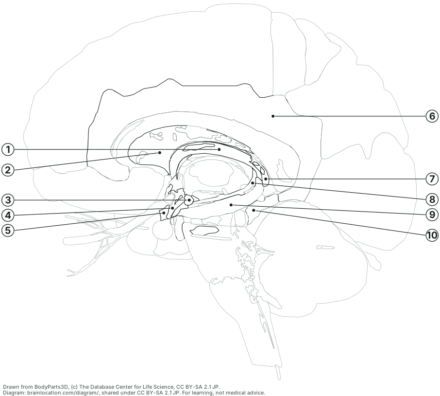
- One half of one adult brain. The right half is drawn; the left half's limbic system mirrors it. The drawing is derived from BodyParts3D and carries its licence, CC BY-SA 2.1 Japan: copy, hand out and reuse it with the credit line. Standalone SVG files: labelled and blank.