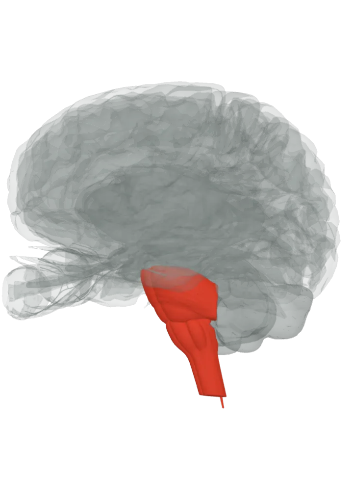
It runs from the reticular formation in the core of the pons and medulla down into the spinal cord, as two tracts: the medial (pontine) and the lateral (medullary) one. They act on the trunk and the upper parts of the limbs, for posture and walking. [1][2]
The reticular formation is not a separate shape in this atlas: steps 1 and 2 light the pons and medulla that hold it; then lit: the parts it runs through, in order.
The brain starts seen from the left, a little from the front and below, where the brainstem runs down to the spinal cord. Step through lights one step at a time, about 1.2 s each, top to bottom; tap a step below to light it. The lit step is in the bright highlight colour, the steps before it stay lit in a paler tint of it and the steps still ahead are grey. Drag to turn, pinch or scroll to zoom. Step through is off because your device asks for reduced motion; tap each step instead.
The tracts and the reticular formation are not drawn: the reticular formation is a diffuse net of cells, not a compact shape, so the first two steps light the whole pons and medulla that hold its two parts, the last the top of the spinal cord, drawn as its thin central canal. [3][1]
The route, in 3 steps
The medial reticulospinal tract, named the pontine tract after where it starts, arises in the caudal and oral pontine reticular nuclei, part of the reticular formation in the core of the pons. Not a separate shape in this atlas; lit: the part that holds it, the pons. [1]Where is the pontine reticular formation (pons)?
The lateral reticulospinal tract, the medullary tract, starts lower down, in the reticular formation of the medulla oblongata, mostly in its gigantocellular (giant-cell) nucleus. Not a separate shape in this atlas; lit: the part that holds it, the medulla oblongata. [1]Where is the medullary reticular formation (medulla)?
Both reach the motor neurons of the trunk and of the flexors and extensors of the upper limbs and hips. The pontine tract ends in laminae VII and VIII of the grey matter; the medullary tract runs the whole length of the cord in the front of the lateral funiculus and ends mostly in lamina VII. [1]Where is the spinal cord?
The two tracts pull in opposite directions on the same muscles: the pontine tract excites the antigravity extensor muscles that hold the body up, the medullary tract inhibits them. [1]
OpenStax describes the reticular system as a diffuse region of grey matter in the brainstem, and says the tract influences the trunk and nearby limb muscles for posture and locomotion, contributes to muscle tone and influences autonomic functions; the medullary tract also takes part in automatic breathing. [2][1]
Two descending systems from the brainstem trigger the automatic postural responses that keep the body balanced and oriented: these tracts from the pons and medulla, and the vestibulospinal tracts from the vestibular nuclei. [1]
It is one of the extrapyramidal tracts, which skip the pyramids of the medulla, with the rubrospinal and tectospinal tracts; by function it joins the vestibulospinal and tectospinal tracts in the medial motor pathway for the trunk and upper limbs rather than the hands. [4][5][2]
The reticular formation also has an ascending part, the reticular activating system, which keeps the cortex awake; this page follows only the descending part. [1]
Facts describe anatomy and normal function only. The 3D model is the adult male BodyParts3D brain (credits and licence); it has no shape for tracts, so a pathway is shown by the parts it runs through.