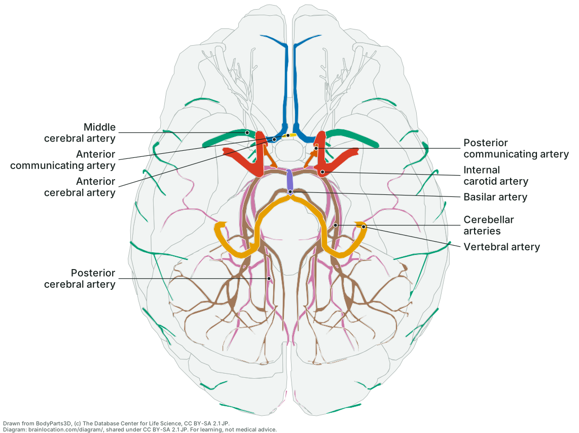
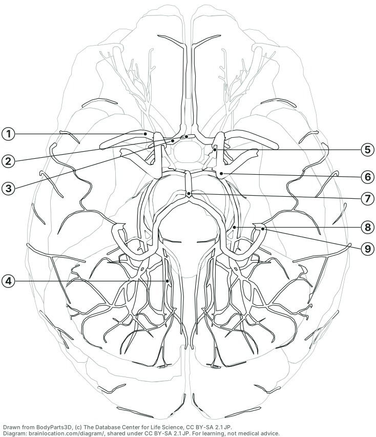
The 9 arteries on the sheet
In the key’s order: the front (carotid) supply, the links, then the back (vertebral–basilar) supply. The same cited lines as the circle of Willis page and each artery’s own page.
- Internal carotid artery (part of the ring): The front source of the ring: each ends under the brain by dividing into the anterior and middle cerebral arteries, after giving off the ophthalmic artery to the eye, the posterior communicating artery and the anterior choroidal artery. (Wikipedia: Internal carotid artery; Wikipedia: Circle of Willis).
- Anterior cerebral artery (part of the ring): Most of the midline (inner) face of the frontal lobe and the upper inner face of the parietal lobe, the front part of the corpus callosum, and deep down the front limb of the internal capsule. (Wikipedia: Anterior cerebral artery).
- Middle cerebral artery: Most of the outer (lateral) surface of each hemisphere, the front of the temporal lobe and the insula; its deep branches feed the basal ganglia and the internal capsule. It leaves the internal carotid but is not itself part of the ring. (Wikipedia: Middle cerebral artery; Wikipedia: Circle of Willis).
- Anterior communicating artery (part of the ring): The front link, about 4 mm long on average, joining the left and right anterior cerebral arteries; it gives off some of the small central branches that feed the optic chiasm, the front of the hypothalamus and the lamina terminalis. (Wikipedia: Anterior communicating artery; Wikipedia: Central arteries).
- Posterior communicating artery (part of the ring): The two side links, each joining an internal carotid to a posterior cerebral artery; they help supply the optic tract and, through small central branches, the front of the thalamus. (Wikipedia: Posterior communicating artery; Wikipedia: Optic tract; Wikipedia: Central arteries).
- Posterior cerebral artery (part of the ring): The occipital lobe and the inner (medial) and under (inferior) faces of the temporal lobe; branches near its start supply a large part of the thalamus. (Wikipedia: Posterior cerebral artery).
- Basilar artery: Together the vertebral arteries and the basilar supply the upper spinal cord, the brainstem, the cerebellum and the back of the brain; the basilar is the main blood supply to the brainstem. (Wikipedia: Vertebral artery).
- Vertebral artery: As the source of the vertebrobasilar system, the vertebral arteries supply the upper spinal cord, the brainstem, the cerebellum and the back of the brain. (Wikipedia: Vertebral artery).
- Cerebellar arteries: On the upper surface of the cerebellum the superior cerebellar artery divides into branches in the pia mater that join those of the two inferior cerebellar arteries. (Wikipedia: Superior cerebellar artery).
How to read it
- You are looking up at the brain from below, front at the top. As on any view from below, the brain’s left half is on the right of the sheet, so the artery on the right of the page is the left one.
- It lies on the underside of the brain, in and around the interpeduncular fossa: the hollow bounded in front by the optic chiasm, with the mammillary bodies and the stalk (infundibulum) of the pituitary gland in its floor. (Wikipedia: Circle of Willis; Wikipedia: Interpeduncular fossa).
- The front of the ring is the short anterior communicating artery, which joins the left and right anterior cerebral arteries in front of the optic chiasm. (Wikipedia: Anterior communicating artery).
- Its sides are the posterior communicating arteries, each running back from the internal carotid to the posterior cerebral artery in the interpeduncular cistern, above and to the side of the pituitary gland. (Wikipedia: Posterior communicating artery).
- The back of the ring is the first part of each posterior cerebral artery, where the basilar artery, lying in the midline on the front of the pons, divides at the junction of the pons and midbrain. (Wikipedia: Basilar artery; Wikipedia: Posterior cerebral artery).
- Which links are thin or missing differs from person to person: the two posterior communicating arteries often differ in size, and in about one adult in five a posterior cerebral artery still draws mainly on the posterior communicating artery rather than the basilar, as it does before birth. (Wikipedia: Posterior communicating artery; Wikipedia: Posterior cerebral artery).
- Its two sources meet in it: the left and right internal carotid arteries, which enter the skull through the carotid canals, and the basilar artery, formed by the two vertebral arteries, which enter through the foramen magnum. The anterior cerebral artery forms the front-side part of the ring, while the middle cerebral artery, which also arises from the internal carotid, leaves next to it and does not contribute to the ring. (OpenStax Anatomy & Physiology 2e, 13.3 Circulation and the Central Nervous System; Wikipedia: Circle of Willis; Wikipedia: Middle cerebral artery).
- Sizes in this model: the basilar artery runs about 28 mm up the front of the pons, and the anterior communicating artery spans about 7 mm from side to side (BodyParts3D 4.0, © DBCLS, CC BY-SA 2.1 Japan); the basilar artery that feeds its back ranges from 1.5 to 6.6 mm across; the two vertebral arteries run about 32 mm inside the skull before they join, and often differ in width: in one study the left was the wider in 54% of people, the right in 30%. (Wikipedia: Basilar artery; Wikipedia: Vertebral artery).
How it is drawn
- The same projection as the underside view, at 1.5 times its scale: every BodyParts3D mesh of the brain and of the blood-supply layer the 3D brain on this site switches on is projected straight up onto the page (no perspective) on a grid of cells. Nothing is traced by hand.
- The arteries are drawn through the brain: where a vessel runs behind a fold of the brain, such as the middle cerebral artery going up into the lateral sulcus, it is shown at its true projected place as if the brain were see-through. Only the parts of the layer up to about 4 mm above the circle’s highest link, the anterior communicating artery, are drawn: higher up, the anterior cerebral arteries climb between the two hemispheres and the middle cerebral arteries spread over their sides, and seen from below those would cover the whole drawing. So the sheet shows the start of each, as an atlas plate of the circle does; the pieces at the brain’s outer edge are middle cerebral branches dipping below that level.
- Which vessel each piece belongs to is read from the data the artery pages are built from, never typed here. The model counts the first stretch of the anterior and of the posterior cerebral arteries in the circle as well; on the sheet each is coloured as its own artery, so the ring is shown as the vessels it is made of.
test/diagram.test.cjsre-projects every artery mesh and fails if a label’s dot is not on its own vessel, or if a label points at another artery’s piece.
What it does not show
- One adult model with a complete ring. Its shape varies between people: the textbook ring is seen in only about one-third of people. (Wikipedia: Circle of Willis). Which links are thin or missing differs from person to person: the two posterior communicating arteries often differ in size, and in about one adult in five a posterior cerebral artery still draws mainly on the posterior communicating artery rather than the basilar, as it does before birth. (Wikipedia: Posterior communicating artery; Wikipedia: Posterior cerebral artery).
- Only the vessels the model has as shapes (BodyParts3D 4.0, © DBCLS, CC BY-SA 2.1 Japan): the ophthalmic, anterior choroidal and small central branches named above are not drawn. The internal carotid and vertebral arteries are cut where the model’s layer stops, at the level of the foramen magnum, and the anterior and middle cerebral arteries where they leave the base. The two vertebral arteries enter the skull through the foramen magnum and join at the lower edge of the pons into the basilar artery; together they supply the upper spinal cord, the brainstem and the cerebellum, and the basilar ends by splitting into the two posterior cerebral arteries. (Wikipedia: Vertebral artery; Wikipedia: Basilar artery; OpenStax Anatomy & Physiology 2e, 13.3 Circulation and the Central Nervous System).
- The drawing is derived from BodyParts3D and carries its licence, CC BY-SA 2.1 Japan: copy, hand out and reuse it with the credit line. Standalone SVG files: labelled and blank. The parts these arteries feed, lit together in 3D: blood supply of the brain.