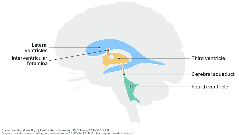
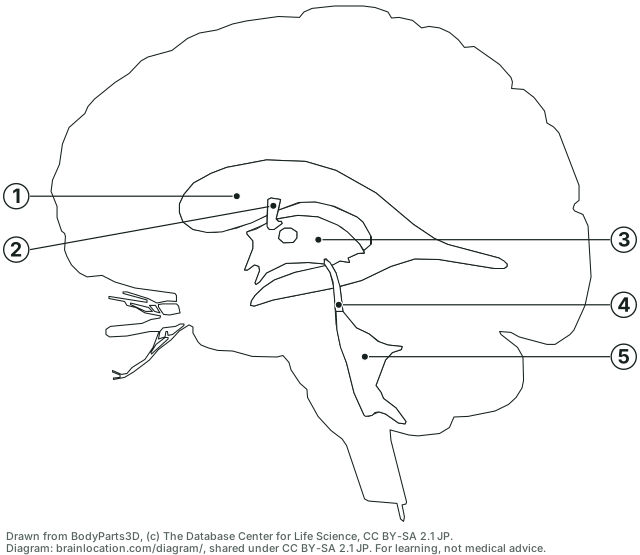
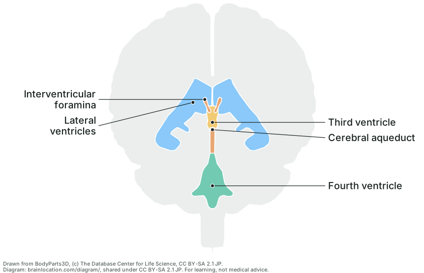
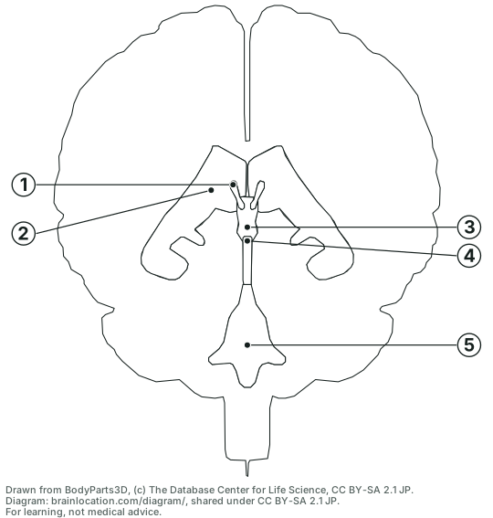
The brain’s fluid-filled spaces on their own, drawn from the 3D model this site uses as if the rest of the brain were glass: from the side and from the front, inside a pale outline of the whole brain so you can see where they sit. Each label links to that part in 3D. Below the labelled sheets are the same two drawings blank and numbered: label them on screen from a pick-list per number and check your answers, or print them with the answer key.
The ventricles from the side
Lateral view of the ventricles inside a see-through brain, seen from the left; front of the brain to the left (the two lateral ventricles lie one behind the other). 5 parts labelled.
Drawn from BodyParts3D, (c) The Database Center for Life Science, CC BY-SA 2.1 JP. Diagram: brainlocation.com/diagram/, shared under CC BY-SA 2.1 JP (credits). For learning, not medical advice.
The ventricles from the front
Anterior view of the ventricles inside a see-through brain, seen from the front; the brain’s left half on the right of the sheet, as when you face someone. 5 parts labelled.
Drawn from BodyParts3D, (c) The Database Center for Life Science, CC BY-SA 2.1 JP. Diagram: brainlocation.com/diagram/, shared under CC BY-SA 2.1 JP (credits). For learning, not medical advice.
The ventricles from the side, blank: name the 5 numbered parts
Lateral view of the ventricles inside a see-through brain, seen from the left; front of the brain to the left (the two lateral ventricles lie one behind the other).
Drawn from BodyParts3D, (c) The Database Center for Life Science, CC BY-SA 2.1 JP. Diagram: brainlocation.com/diagram/, shared under CC BY-SA 2.1 JP (credits). For learning, not medical advice.
The ventricles from the front, blank: name the 5 numbered parts
Anterior view of the ventricles inside a see-through brain, seen from the front; the brain’s left half on the right of the sheet, as when you face someone.
Drawn from BodyParts3D, (c) The Database Center for Life Science, CC BY-SA 2.1 JP. Diagram: brainlocation.com/diagram/, shared under CC BY-SA 2.1 JP (credits). For learning, not medical advice.
Download the SVG files
Vector files that scale to any size without blurring, on a white background with the credit line built in: open one in a browser, an office suite or a drawing program, or drop it into slides and handouts.
On the side view the C shape of the lateral ventricle is easy to see: it starts in the frontal lobe (the left end of the drawing), runs back over the thalamus, and curls down and forward into the temporal lobe. The left and right lateral ventricles lie one behind the other here, so they draw as one shape. On the front view they separate into two, one either side of the midline, with the thin third ventricle standing between them below.
The small round gap through the third ventricle on the side view is real anatomy, not a flaw of the drawing: there the left and right thalamus touch across the ventricle, the interthalamic adhesion (massa intermedia), which not every brain has (Wikipedia: Interthalamic adhesion; Wikipedia: Third ventricle).
Measured on the model: one lateral ventricle runs about 109 mm from front to back and 49 mm from top to bottom; the third ventricle is about 8 mm across at its widest but 40 mm long; the fourth ventricle is about 28 mm wide; the aqueduct about 18 mm tall and 3 mm across. Real brains differ, and the ventricles differ more than most parts.
How it is drawn
The shapes are not traced by hand: the BodyParts3D meshes the 3D brain on this site shows are projected straight onto the page (no perspective) on a grid of 0.25 mm cells. The pale shape is the outline of every brain part together. The five ventricle parts are drawn as if the brain were see-through; where two overlap on the page the smaller channel is drawn on top (the foramina, then the aqueduct, the third ventricle, the fourth and last the lateral ventricles), so each keeps a visible shape.
Each label starts at the point deepest inside its own shape: test/diagram.test.cjs checks that every dot lies inside the outline drawn for that part and that a line of sight through the dot really passes through that part’s 3D mesh.
What it does not show
The horns of the lateral ventricle (the frontal or anterior horn, the body, the atrium where it bends, the occipital or posterior horn and the temporal or inferior horn) are not separate shapes in the model, so they are not labelled; each lateral ventricle is one shape here (Wikipedia: Lateral ventricles).
The choroid plexus, the tissue that makes most of the fluid, lines the body, atrium and temporal horn of each lateral ventricle and the roofs of the third and fourth ventricles (Wikipedia: Choroid plexus; Wikipedia: Ventricular system). It is tissue inside the spaces, not a space, so it is left off this sheet; the model has a shape for it only in the lateral ventricles.
The three openings through which the fluid leaves the fourth ventricle (one median, two lateral apertures) and the central canal of the spinal cord below it have no shapes in the model, so they are not drawn (Wikipedia: Ventricular system).
It is one adult brain. The drawing is derived from BodyParts3D and carries its licence, CC BY-SA 2.1 Japan: copy, hand out and reuse it with the credit line. Standalone SVG files: side labelled and blank, front labelled and blank.