Front page › All parts of the brain › Sizes
How big is the brain, and each part in it
On the one adult brain model this site is built on, the whole brain fits in a box about 180 mm front to back, 141 mm side to side and 166 mm top to bottom [1]. A typical adult brain weighs about 1.2–1.4 kg [2].
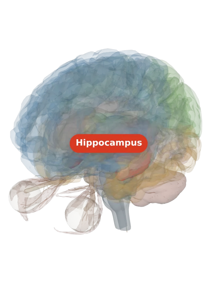
Loading the 3D brain…
Lit: the hippocampus, about 43 × 20 × 22 mm. Tap “Light it” on any row.
Drag to turn, pinch or scroll to zoom, tap any part to name it. A small part deep inside shows best with a slice: try Side or Front after lighting it.
Each “Light it” button lights exactly what that part’s own page lights, and the caption under the brain gives its box in millimetres: length (front to back) × width (side to side) × height (top to bottom).
Every part with a shape, largest first
Measured from the BodyParts3D model, one adult. Each row is the box the part fits in, in millimetres, exactly as its own page gives it: length front to back, width side to side, height top to bottom. The bar under each name is its longest side against the whole brain’s 180 mm.
| Part | Length mm | Width mm | Height mm | Light it |
|---|---|---|---|---|
| Cerebrum all its parts together, both sides | 180 | 141 | 117 | |
| Forebrain all its parts together, both sides | 180 | 141 | 117 | |
| White matter white matter of both hemispheres with the corpus callosum | 163 | 125 | 100 | |
| Inferior parietal lobule all its parts together, both sides | 74 | 139 | 64 | |
| Frontal lobe the left one; the right is its mirror image | 130 | 63 | 97 | |
| Prefrontal cortex the left one; the right is its mirror image | 116 | 61 | 97 | |
| Superior frontal gyrus the left one; the right is its mirror image | 116 | 32 | 78 | |
| Temporal lobe the left one; the right is its mirror image | 114 | 61 | 60 | |
| Limbic system all its parts together, both sides | 111 | 75 | 76 | |
| Cingulate gyrus the left one; the right is its mirror image | 111 | 28 | 67 | |
| Middle temporal gyrus the left one; the right is its mirror image | 111 | 36 | 34 | |
| Ventricles of the brain all its parts together, both sides | 109 | 78 | 88 | |
| Lateral ventricles the left one; the right is its mirror image | 109 | 39 | 49 | |
| Hindbrain all its parts together | 85 | 106 | 76 | |
| Cerebellum | 56 | 106 | 53 | |
| Superior temporal gyrus the left one; the right is its mirror image | 106 | 43 | 49 | |
| Inferior temporal gyrus the left one; the right is its mirror image | 96 | 44 | 29 | |
| Parietal lobe the left one; the right is its mirror image | 94 | 69 | 84 | |
| Corpus callosum | 92 | 51 | 39 | |
| Tentorium cerebelli right half of the tentorium cerebelli (the only half the atlas ships) | 87 | 60 | 33 | |
| Middle frontal gyrus the left one; the right is its mirror image | 85 | 35 | 62 | |
| Brainstem | 46 | 42 | 81 | |
| Trochlear nerve the left one; the right is its mirror image | 79 | 9 | 12 | |
| Basal ganglia all its parts together, both sides | 64 | 70 | 37 | |
| Postcentral gyrus the left one; the right is its mirror image | 64 | 67 | 60 | |
| Superior parietal lobule the left one; the right is its mirror image | 57 | 36 | 67 | |
| Orbitofrontal cortex the left one; the right is its mirror image | 67 | 40 | 30 | |
| Precentral gyrus the left one; the right is its mirror image | 66 | 62 | 66 | |
| Caudate nucleus the left one; the right is its mirror image | 64 | 32 | 37 | |
| Occipital lobe the left one; the right is its mirror image | 63 | 44 | 54 | |
| Inferior frontal gyrus the left one; the right is its mirror image | 63 | 35 | 60 | |
| Angular gyrus the left one; the right is its mirror image | 46 | 43 | 63 | |
| Septum (septum of telencephalon) | 62 | 20 | 23 | |
| Fusiform gyrus the left one; the right is its mirror image | 61 | 32 | 27 | |
| Internal capsule the left one; the right is its mirror image | 54 | 23 | 33 | |
| Insula the left one; the right is its mirror image | 52 | 19 | 35 | |
| Diencephalon all its parts together, both sides | 51 | 48 | 34 | |
| Optic nerve the left one; the right is its mirror image | 49 | 32 | 22 | |
| Commissure of the fornix | 31 | 49 | 20 | |
| Parahippocampal gyrus the left one; the right is its mirror image | 49 | 21 | 22 | |
| Extraocular muscles the left one; the right is its mirror image | 47 | 32 | 31 | |
| Supramarginal gyrus the left one; the right is its mirror image | 38 | 46 | 42 | |
| Fornix the left one; the right is its mirror image | 45 | 31 | 32 | |
| Epithalamus all its parts together, both sides | 44 | 15 | 21 | |
| Choroid plexus the left one; the right is its mirror image | 43 | 37 | 34 | |
| Stria terminalis the left one; the right is its mirror image | 43 | 25 | 31 | |
| Putamen the left one; the right is its mirror image | 43 | 18 | 26 | |
| Hippocampus the left one; the right is its mirror image | 43 | 20 | 22 | |
| Pons | 34 | 42 | 29 | |
| Medulla oblongata | 33 | 27 | 41 | |
| Third ventricle | 40 | 8 | 23 | |
| Fourth ventricle | 23 | 28 | 38 | |
| Optic tract the left one; the right is its mirror image | 38 | 25 | 8 | |
| Thalamus the left one; the right is its mirror image | 37 | 24 | 24 | |
| Stria medullaris the left one; the right is its mirror image | 37 | 4 | 16 | |
| Oculomotor nerve the left one; the right is its mirror image | 36 | 25 | 30 | |
| Spinal cord | 13 | 2 | 36 | |
| Midbrain | 29 | 29 | 17 | |
| Globus pallidus the left one; the right is its mirror image | 29 | 17 | 17 | |
| Eyeball the left one; the right is its mirror image | 25 | 26 | 26 | |
| Anterior commissure | 5 | 24 | 3 | |
| Cerebral peduncles the left one; the right is its mirror image | 23 | 19 | 17 | |
| Hypothalamus | 18 | 17 | 20 | |
| Amygdala the left one; the right is its mirror image | 18 | 17 | 13 | |
| Cerebral aqueduct | 7 | 3 | 18 | |
| Optic chiasm | 10 | 15 | 8 | |
| Posterior commissure | 5 | 15 | 3 | |
| Pituitary gland | 10 | 12 | 13 | |
| Interventricular foramina one shape the atlas draws for both interventricular foramina | 5 | 13 | 11 | |
| Lamina terminalis lamina terminalis (both halves the atlas draws) | 7 | 3 | 12 | |
| Lateral geniculate body the left one; the right is its mirror image | 11 | 8 | 7 | |
| Pineal gland | 11 | 6 | 8 | |
| Inferior colliculus the left one; the right is its mirror image | 8 | 8 | 8 | |
| Medial geniculate body the left one; the right is its mirror image | 7 | 7 | 8 | |
| Superior colliculus the left one; the right is its mirror image | 6 | 8 | 6 | |
| Habenula | 5 | 8 | 4 | |
| Mammillary bodies the left one; the right is its mirror image | 5 | 4 | 5 |
How to read these numbers
- One ruler. Every row is measured the same way on the same model: the smallest box, lined up with the head’s front–back, side–side and up–down axes, that holds every point of the part’s surface, rounded to the millimetre. So rows compare fairly with each other: on this model the cerebrum is 4.2 times as long, on its longest side, as the hippocampus, and the mammillary bodies is the smallest row at 5 × 4 × 5 mm.
- A box, not a volume. A curved, slanted or branching part fills only some of its box: the fornix arches up and over the thalamus, so its box holds a lot of space that is not fornix. Length × width × height therefore overstates any part’s volume, and this page gives no volumes because it does not check that the model’s surfaces are closed, which a volume needs.
- Paired parts are measured by their left copy; the right one is its mirror image on this model. Groups (the basal ganglia, the ventricles, the forebrain) are the one box all their members fit in together.
- One adult, not everyone. The model is one adult male (BodyParts3D). Real brains differ from person to person, as the weight range above shows, so these are the sizes of one brain, not a standard and not a yardstick for anyone’s brain.
Pages not in the table, and why
- 47 parts too small for the atlas to draw (the substantia nigra, for one). The model has no shape for them, so their pages light and measure the larger part that holds them; that number is the holder’s size, not theirs, and listing it here would pass it off as theirs.
- 24 areas and named parts that the model draws only as part of a larger shape (Broca’s area, for one). Each of their pages measures the whole shape that holds it, which is not the area’s own size.
- 14 grooves (the central sulcus, for one). A groove is the gap between two shapes, not a shape; its page measures where its two banks meet.
- 8 cranial nerves the model has no shape for (the vagus nerve, for one). Their pages light and measure the part each leaves the brain from.
- 10 artery pages (the circle of Willis, for one). An artery’s box says how far its branches spread over the brain, not how big the vessel is; each artery page gives that spread, and the blood supply page shows them together.
- The cerebral hemispheres: their page measures the left and right halves as two boxes; the cerebrum row here is both together.
Sources
- BodyParts3D 4.0 (3D model measured on this page), © DBCLS, CC BY-SA 2.1 Japan
- Wikipedia: Human brain
Facts describe anatomy and normal function only. The 3D model is the adult male BodyParts3D brain (credits and licence); real brains vary in size and shape, so its measurements describe one model, not anyone’s brain.